Computer Vision Problems

Computer vision problems or Digital eye strain is a group of eye and vision problems. The problems can include eyes that itch and tear, and are dry and red. Your eyes may feel tired or uncomfortable. You may not be able to focus normally. These problems are caused by lots of computer or digital device use. Using e-readers and smartphones may also cause these problems.

These problems have been increasing in frequency over the past few decades. Many people have some symptoms if they use a computer or digital device for long periods. Most computer or digital device users have symptoms at least some of the time. Digital eye strain is very common in both children and adults.

What causes digital eye strain?

For many reasons, reading text on a computer screen or digital device is often harder for the eyes than reading printed text. This is why working on a computer for a few hours may cause symptoms of digital eye strain, but reading a book may not.

Several factors help to cause digital eye strain, such as:

  • Screen glare
  • Poor lighting
  • Poor posture while using a computer
  • Viewing a computer at the wrong distance and angle
  • Uncorrected vision problems
  • A combination of many of these factors

People often blink less when using a computer than when reading printed text. This can cause dry eye, This may contribute to digital eye strain.

Who is at risk for getting digital eye strain?

You may be at greater risk for digital eye strain if you:

  • Spend a few hours or more a day at a computer or on a digital device
  • Are too close to your computer or digital device screen
  • View your computer or digital device at the wrong angle
  • Have bad posture while using your computer or digital device
  • Have eye problems (even minor ones) not corrected with glasses or contact lenses
  • Have a pair of glasses that is not suitable for viewing the distance of your computer
  • Don’t take breaks while you are working

You may have an underlying problem with dry eye. This may make digital eye strain worse, or more likely to occur. Dry eye is more common in women than in men. It also becomes more common with age. Some medicines and health problems make dry eye more likely. For example, if you use antihistamines, you may be at greater risk of having dry eye. If you have thyroid disease or certain autoimmune diseases, you are also at greater risk of having dry eye.

What are the symptoms of digital eye strain?

Digital eye strain can cause many symptoms, including:

  • Blurred vision
  • Double vision
  • Dry eye
  • Eye discomfort
  • Eye fatigue
  • Eye itching
  • Eye redness
  • Eye tearing
  • Headaches
  • Neck and shoulder pain

Most of these symptoms are short-term (temporary). They often lessen or go away when you stop using your computer or device. But symptoms may continue for a longer time.

The severity of symptoms may vary depending on:

  • How long you have been using the computer or digital device
  • Your underlying eye problems
  • Other factors that cause digital eye strain.

Symptoms may get worse if you don’t resolve the problem.

Using a computer or digital device for a long time can also lead to other symptoms. This includes neck and shoulder pain. This is often due to poor alignment and posture when using the computer or digital device. Some healthcare providers call these symptoms part of digital eye strain as well.

How is digital eye strain diagnosed?

Your eye care provider will make a diagnosis with a health history and complete eye exam. They will assess if any health problems, medicines, or environmental factors might be adding to your symptoms.

Your eye care provider may test the sharpness of your vision and how well your eyes focus and work together. For a more detailed exam your provider may want to dilate (enlarge) your pupils. Then they will use a device (ophthalmoscope) to look at the back of your eye. In some cases, you may need to get follow-up blood tests for healthcare problems that might be helping to cause your digital eye strain.

How is digital eye strain treated?

Treatment includes creating a better work environment.

  • Rest your eyes at least 15 minutes after each 2 hours of computer or digital device use.
  • Every 20 minutes, look into the distance at least 20 feet away from the computer or digital device. Do this for at least 20 seconds.
  • Enlarge the text on your computer screen or digital device.
  • Reduce glare from the light sources in your environment.
  • Think about using a screen glare filter.
  • Place your screen so that the center of it is about 4 to 5 inches below eye level (about 15 to 20 degrees from the horizontal).
  • Place your screen about 20 to 28 inches from your eye.( About arm’s length.)
  • Remember to blink often.
  • Fix your chair height so your feet can rest comfortably on the floor. Don’t slump over the computer screen.

Making these changes may help eliminate digital eye strain in many people.

Your eye care provider will also need to treat any hidden health problems that may be adding to your digital eye strain. For instance, you might need a new pair of glasses. If you have an underlying dry eye problem, your eye care provider might advise the following:

  • Using lubricating drops
  • Treating allergies, if you have them
  • Creating a more humid work environment
  • Drinking more fluids (staying hydrated)
  • Taking a prescription medicine to increase tear production

What can I do to prevent digital eye strain?

Create a better work environment to help prevent digital eye strain. If you use glasses or corrective lenses, see your eye care provider at least once a year or as advised for a checkup. Also see your healthcare provider regularly. This can help prevent and treat health problems that can help cause digital eye strain.

Key points about digital eye strain

  • Digital eye strain is a group of related eye and vision problems caused by extended computer or digital device use.
  • Symptoms include eye discomfort and fatigue, dry eye, blurry vision, and headaches.
  • Uncorrected vision problems are a major cause.
  • Sometimes hidden health problems help to cause it.
  • Having a better computer work environment may help improve symptoms.
  • Resting your eyes regularly is one of the best ways to prevent and treat digital eye strain.

Next steps

Tips to help you get the most from a visit to your healthcare provider:

  • Know the reason for your visit and what you want to happen.
  • Before your visit, write down questions you want answered.
  • Bring someone with you to help you ask questions and remember what your healthcare provider tells you.
  • At the visit, write down the name of a new diagnosis, and any new medicines, treatments, or tests. Also write down any new instructions your healthcare provider gives you.
  • Know why a new medicine or treatment is prescribed, and how it will help you. Also know what the side effects are.
  • Ask if your condition can be treated in other ways.
  • Know why a test or procedure is recommended and what the results could mean.
  • Know what to expect if you do not take the medicine or have the test or procedure.
  • If you have a follow-up appointment, write down the date, time, and purpose for that visit.
  • Know how you can contact your healthcare provider if you have questions.

Headaches and your eyes

How a Headache Can Affect Your Eyes and Vision

By Troy Bedinghaus, OD  Medically reviewed by Diana Apetauerova, MD on September 08, 2020

Have you ever had a headache that affected your vision? Sometimes a headache can cause pain around your eyes, even though the headache is not associated with a vision problem. On the other hand, a headache may be a sign that your eyes are changing and that it’s time to schedule an eye exam. Although headaches are rarely a medical emergency, a severe one should not be ignored or minimized.

headaches and vision
Verywell / Luyi Wang

Headaches That Affect Vision

Vision problems can sometimes be the consequence of a headache. This is especially true with migraines and cluster headaches.

Migraine Headache

A migraine headache can cause intense pain in and around your eyes. A migraine aura resembling flashing lights, a prismatic rainbow of lights or a zig-zag pattern of shimmering lights often precedes the actual headache. The aura typically lasts around 20 minutes.

Some people who experience a migraine aura never develop the actual headache, making the diagnosis of the visual disturbances difficult.1 Migraines can also cause tingling or numbness of the skin. People with severe migraines may experience nausea, vomiting, and light sensitivity. Medications, certain foods, smells, loud noises, and bright lights can all trigger a migraine headache.An Overview of Migraine With Aura

Cluster Headache

Cluster headaches are severe headaches that occur in clusters and typically cause pain around the eyes. The pain often radiates down the neck to include the shoulder. Other symptoms include:

Cluster headaches may occur daily for several months at a time followed by a long period with no headaches. It is not known what causes cluster headaches, but they are clearly one of the most severe headaches one can experience.

Vision Problems That Cause Headaches

On the flip side, vision problems can cause headaches when you either overwork the eyes or struggle to maintain focus. By correcting the vision problem, you can often resolve the headache.

Eye Strain

Simply overusing the focusing muscles of your eyes can cause eye strain and headaches. This is an increasing problem in our high tech world

Small-screen texting and web browsing can easily cause eye strain, in part because the words and images on a computer screen are made up of pixels and do not have well-defined edges. The eyes cannot easily focus on pixels, so they must work harder even if an image is in high-resolution.2 When the eye muscles become fatigued, a headache can develop around or behind the eyes.

Farsightedness

Adults and children with uncorrected farsightedness (hypermetropia) will often experience a frontal headache (also known as a “brow ache”). If you are farsighted, you may find it difficult to focus on nearby objects, resulting in eye strain and headaches. As you subconsciously compensate for your farsightedness by focusing harder, the headaches can become worse and more frequent.

Presbyopia

Around the age of 40, people begin to find it difficult to focus on nearby objects. Near point activities, such as reading or threading a needle, are often difficult to perform because of blurring. This is an unavoidable condition known as presbyopia that affects everyone at some point. Headaches develop as you try to compensate for the lack of focusing power. Reading glasses can often relieve the underlying eye strain.

Occupations requiring close-up work, exposure to sunlight for longer periods of time, and farsightedness were the most common risk factors for presbyopia.3Presbyopia: Close-Up Vision Loss and What to Do About It

Giant Cell Arteritis

Also known as temporal arteritis, giant cell arteritis (GCA) is an inflammation of the lining of the arteries that run along the temple. GCA usually creates a headache that causes constant, throbbing pain in the temples. Vision symptoms occur as a result of a loss of blood supply to the optic nerve and retina. Other symptoms include:

  • Fever, fatigue and muscle aches
  • Scalp tenderness
  • Pain while chewing
  • Decreased vision​

GCA is considered a medical emergency. If left untreated, the condition may cause vision loss in one or both eyes. A delayed diagnosis is the most common cause of GCA-associated vision loss.42:18

What Is a Retinal Migraine?

Acute Angle-Closure Glaucoma 

Acute angle-closure glaucoma (AACG) is a rare type of glaucoma that causes a sudden onset of symptoms, including headaches. Eye pressure rises quickly in AACG causing increased eye redness, eye pain, and cloudy vision. A mid-dilated pupil (in which pupil dilation is sluggish and incomplete) is one of the most important diagnostic features of AACG.5

Ocular Ischemic Syndrome

Ocular ischemic syndrome (OIS) is a condition that develops due to a chronic lack of blood flow to the eye. This condition often causes a headache, decreased vision, and a host of other signs, including cataracts, glaucoma, iris neovascularization (the development of new weak blood vessels in the iris), and retinal hemorrhage. White spots on the retina indicate a lack of blood flow and oxygen to the retinal tissue.2:18

What Is a Retinal Migraine?

Herpes Zoster

Also known as shingles, herpes zoster is known for causing headaches, vision changes and severe pain around the head and eye. Herpes zoster is a reactivation of the chickenpox virus and affects a single side of the body. A headache usually precedes an outbreak of painful skin blisters.

Herpes zoster around the eyes is serious and requires immediate medical attention (including antiviral medication) to prevent damage to the ocular nerves and eyes. Complications include corneal clouding, glaucoma, and optic nerve atrophy (deterioration).6

Pseudotumor Cerebri

Pseudotumor cerebri is a condition that occurs when the pressure within the skull increases for no apparent reason. For this reason, pseudotumor cerebri is also referred to as Idiopathic Intracranial hypertension (“idiopathic” meaning of unknown origin and “hypertension” meaning high blood pressure).

Pseudotumor cerebri often causes a headache and changes in vision. If left untreated, pseudotumor cerebri can lead to vision loss as the pressure places strain on the optic nerves. Fortunately, while 65% to 85% of people with pseudotumor cerebri will experience visual impairment, the condition is usually transient and will normalize when the hypertension is controlled.

Don’t let glaucoma steal your vision

Overview

Open-angle glaucomaOpen-angle glaucomaOpen pop-up dialog box
Glaucoma is a group of eye conditions that damage the optic nerve, the health of which is vital for good vision. This damage is often caused by an abnormally high pressure in your eye.

Glaucoma is one of the leading causes of blindness for people over the age of 60. It can occur at any age but is more common in older adults.

Many forms of glaucoma have no warning signs. The effect is so gradual that you may not notice a change in vision until the condition is at an advanced stage.

Because vision loss due to glaucoma can’t be recovered, it’s important to have regular eye exams that include measurements of your eye pressure so a diagnosis can be made in its early stages and treated appropriately. If glaucoma is recognized early, vision loss can be slowed or prevented. If you have the condition, you’ll generally need treatment for the rest of your life.

Symptoms

The signs and symptoms of glaucoma vary depending on the type and stage of your condition. For example:

Open-angle glaucoma

  • Patchy blind spots in your side (peripheral) or central vision, frequently in both eyes
  • Tunnel vision in the advanced stages

Acute angle-closure glaucoma

  • Severe headache
  • Eye pain
  • Nausea and vomiting
  • Blurred vision
  • Halos around lights
  • Eye redness

If left untreated, glaucoma will eventually cause blindness. Even with treatment, about 15 percent of people with glaucoma become blind in at least one eye within 20 years.

REQUEST AN EYE EXAMINATION AT EYEUPDATE CLINIC

When to see a doctor

Promptly go to an emergency room or an eye doctor’s (ophthalmologist’s) office if you experience some of the symptoms of acute angle-closure glaucoma, such as severe headache, eye pain and blurred vision.

Causes

Glaucoma is the result of damage to the optic nerve. As this nerve gradually deteriorates, blind spots develop in your visual field. For reasons that doctors don’t fully understand, this nerve damage is usually related to increased pressure in the eye.

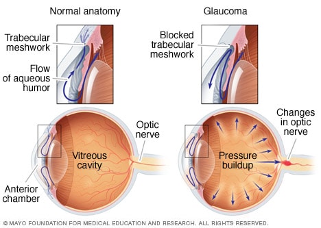
Elevated eye pressure is due to a buildup of a fluid (aqueous humor) that flows throughout the inside of your eye. This internal fluid normally drains out through a tissue called the trabecular meshwork at the angle where the iris and cornea meet. When fluid is overproduced or the drainage system doesn’t work properly, the fluid can’t flow out at its normal rate and eye pressure increases.

Glaucoma tends to run in families. In some people, scientists have identified genes related to high eye pressure and optic nerve damage.

Types of glaucoma include:

Open-angle glaucoma

Open-angle glaucoma is the most common form of the disease. The drainage angle formed by the cornea and iris remains open, but the trabecular meshwork is partially blocked. This causes pressure in the eye to gradually increase. This pressure damages the optic nerve. It happens so slowly that you may lose vision before you’re even aware of a problem.

Angle-closure glaucoma

Angle-closure glaucoma, also called closed-angle glaucoma, occurs when the iris bulges forward to narrow or block the drainage angle formed by the cornea and iris. As a result, fluid can’t circulate through the eye and pressure increases. Some people have narrow drainage angles, putting them at increased risk of angle-closure glaucoma.

Angle-closure glaucoma may occur suddenly (acute angle-closure glaucoma) or gradually (chronic angle-closure glaucoma). Acute angle-closure glaucoma is a medical emergency.

Normal-tension glaucoma

In normal-tension glaucoma, your optic nerve becomes damaged even though your eye pressure is within the normal range. No one knows the exact reason for this. You may have a sensitive optic nerve, or you may have less blood being supplied to your optic nerve. This limited blood flow could be caused by atherosclerosis — the buildup of fatty deposits (plaque) in the arteries — or other conditions that impair circulation.

Glaucoma in children

It’s possible for infants and children to have glaucoma. It may be present from birth or develop in the first few years of life. The optic nerve damage may be caused by drainage blockages or an underlying medical condition.

Pigmentary glaucoma

In pigmentary glaucoma, pigment granules from your iris build up in the drainage channels, slowing or blocking fluid exiting your eye. Activities such as jogging sometimes stir up the pigment granules, depositing them on the trabecular meshwork and causing intermittent pressure elevations.

REQUEST AN EYE EXAMINATION AT EYEUPDATE CLINIC

Risk factors

Because chronic forms of glaucoma can destroy vision before any signs or symptoms are apparent, be aware of these risk factors:

  • Having high internal eye pressure (intraocular pressure)
  • Being over age 60
  • Being black, Asian or Hispanic
  • Having a family history of glaucoma
  • Having certain medical conditions, such as diabetes, heart disease, high blood pressure and sickle cell anemia
  • Having corneas that are thin in the center
  • Being extremely nearsighted or farsighted
  • Having had an eye injury or certain types of eye surgery
  • Taking corticosteroid medications, especially eyedrops, for a long time

Prevention

These self-care steps can help you detect glaucoma in its early stages, which is important in preventing vision loss or slowing its progress.

  • Get regular dilated eye examinations. Regular comprehensive eye exams can help detect glaucoma in its early stages, before significant damage occurs. As a general rule, the American Academy of Ophthalmology recommends having a comprehensive eye exam every five to 10 years if you’re under 40 years old; every two to four years if you’re 40 to 54 years old; every one to three years if you’re 55 to 64 years old; and every one to two years if you’re older than 65. If you’re at risk of glaucoma, you’ll need more frequent screening. Ask your doctor to recommend the right screening schedule for you.
  • Know your family’s eye health history. Glaucoma tends to run in families. If you’re at increased risk, you may need more frequent screening.
  • Exercise safely. Regular, moderate exercise may help prevent glaucoma by reducing eye pressure. Talk with your doctor about an appropriate exercise program.
  • Take prescribed eyedrops regularly. Glaucoma eyedrops can significantly reduce the risk that high eye pressure will progress to glaucoma. To be effective, eyedrops prescribed by your doctor need to be used regularly even if you have no symptoms.
  • Wear eye protection. Serious eye injuries can lead to glaucoma. Wear eye protection when using power tools or playing high-speed racket sports in enclosed courts.

Have an eye check within Omole Akiode Ojodu Berger for detection of Age Related Macular Degeneration (ARMD)

Age-related macular degeneration (AMD) is the most common cause of irreversible central vision loss in older patients. Dilated funduscopic findings are diagnostic; color photographs, fluorescein angiography, and optical coherence tomography assist in confirming the diagnosis and in directing treatment. Treatment is with dietary supplements, intravitreal injection of antivascular endothelial growth factor drugs, laser photocoagulation, photodynamic therapy, and low-vision devices.

Risk factors include the following:

  • Age

  • Genetic variants (eg, abnormal complement factor H)

  • Family history

  • Smoking

  • Obesity

  • Sun exposure

  • A diet low in omega-3 fatty acids and dark green leafy vegetables

    Age-Related Macular Degeneration (AMD or ARMD)

    (Senile Macular Degeneration)

    By

    Sonia Mehta

    , MD, Vitreoretinal Diseases and Surgery Service, Wills Eye Hospital, Sidney Kimmel Medical College at Thomas Jefferson University

    Professional.Manuals.TopicPage.LastRevisionDate| Content last modified Jun 2019
    CLICK HERE FOR PATIENT EDUCATION


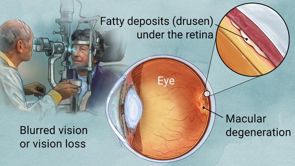
    Age-related macular degeneration (AMD) is the most common cause of irreversible central vision loss in older patients. Dilated funduscopic findings are diagnostic; color photographs, fluorescein angiography, and optical coherence tomography assist in confirming the diagnosis and in directing treatment. Treatment is with dietary supplements, intravitreal injection of antivascular endothelial growth factor drugs, laser photocoagulation, photodynamic therapy, and low-vision devices.


    AMD is the leading cause of permanent, irreversible vision loss in older adults. It is more common among whites.


    Two different forms occur:

    • Dry (nonexudative or atrophic): All AMD starts as the dry form. About 85% of people with AMD have only dry AMD.

    • Wet (exudative or neovascular): Wet AMD occurs in about 15% of people.

    Normal Retina
    Normal Retina
    IMAGE PROVIDED BY SUNIR GARG, MD.

Contact us

Eye clinic locations:

Eye centre Akute Ajuwon Fagba Alagbole Agege Alagbado Matogun Olambe Lambe Obawole Giwa Okearo abule Egba Ogba

01, Ajuwon junction, Ajuwon bus stop, beside BPNL filling station, Akute/Ajuwon road, off Elliot bus stop, Iju-Ishagah. Tel: 08034971582

222, Iju road, Balogun, Ifako Ijaye, Lagos. Tel: 08107531046

01, Itshekiri street, beside M. R. S. Petrol station, Ajuwon, Ifo, Ogun state. Tel: 08068084591.

Astigmatism

According to American Optometric Association (AOA), Astigmatism is a common vision condition that causes blurred vision. It occurs when the cornea (the clear front cover of the eye) is irregularly shaped or sometimes because of the curvature of the lens inside the eye.

An irregularly shaped cornea or lens prevents light from focusing properly on the retina, the light-sensitive surface at the back of the eye. As a result, vision becomes blurred at any distance. This can lead to eye discomfort and headaches.

Astigmatism frequently occurs with other vision conditions like myopia (nearsightedness) and hyperopia (farsightedness). Together these vision conditions are referred to as refractive errors because they affect how the eyes bend or “refract” light.

There are many causes to astigmatism. It can be hereditary and is usually present from birth. It can decrease or increase over time.

A comprehensive optometric examination will include testing for astigmatism. If necessary, your optometrist can provide eyeglasses or contact lenses that correct the astigmatism by altering the way light enters the eyes.

Another option for treating astigmatism is a corneal procedure called orthokeratology (ortho-k). In this painless, noninvasive procedure, the patient wears a series of specially designed rigid contact lenses to gradually reshape the curvature of the cornea.

Laser surgery can also treat some types of astigmatism. The laser changes the shape of the cornea by removing a small amount of eye tissue.

[back to top]

What causes astigmatism?

The curvature of the cornea and lens bends the light entering the eye in order to focus it precisely on the retina at the back of the eye. In astigmatism, the surface of the cornea or lens has a somewhat different curvature.

the surface of the cornea is shaped more like a football instead of round like a basketball, the eye is unable to focus light rays to a single point. Vision becomes out of focus at any distance.

In addition, the curvature of the lens inside the eye can change, resulting in an increase or decrease in astigmatism. This change frequently occurs in adulthood and can precede the development of naturally occurring cataracts.

Sometimes astigmatism may develop following an eye injury or eye surgery.

Astigmatism also occurs due to a relatively rare condition called keratoconus in which the cornea becomes progressively thinner and cone-shaped. This results in a large amount of astigmatism, which causes poor vision that cannot be clearly corrected with eyeglasses. People with keratoconus usually need contact lenses for clear vision and eventually may need a corneal transplant.

An optometrist can diagnose an astigmatism through a comprehensive eye examination. Testing for astigmatism measures how the eyes focus light and determines the power of any optical lenses needed to improve vision. This examination may include:

Visual acuity-When you read letters on a distance chart, you are measuring your visual acuity. Visual acuity is given as a fraction (for example, 20/40). The top number is the standardized testing distance (20 feet) and the bottom number is the smallest letter size read. A person with 20/40 visual acuity would have to get within 20 feet to read a letter that should be seen clearly at 40 feet. Normal distance visual acuity is 20/20.

Keratometry/Topography-A keratometer is the primary instrument used to measure the curvature of the cornea. By focusing a circle of light on the cornea and measuring its reflection, it is possible to determine the exact curvature of that area of the cornea’s surface. This measurement is particularly critical in determining the proper fit for contact lenses. A corneal topographer, which is gaining use, generates a contour map of the cornea and provides even more detail of the cornea’s shape.

Refraction-Using an instrument called a phoropter, your optometrist places a series of lenses in front of your eyes and measures how they focus light. This is performed using a handheld lighted instrument called a retinoscope or an automated instrument that evaluates the approximate focusing power of the eye. Based on your responses, the power is then refined to determine the lenses that allow the clearest vision. Despite improved technology, patient input remains integral in determining vision needs.
With the information from these tests, your optometrist can determine if you have astigmatism. Your optometrist will use these findings, combined with those of other tests performed, to determine the power of any lens correction you need to provide clear, comfortable vision. Once testing is complete, your optometrist can discuss treatment options.

[back to top]

How is astigmatism treated?

People with astigmatism have several options to regain clear vision. They include:

Eyeglasses. People with astigmatism primarily choose eyeglasses to improve their vision. The eyeglasses contain a special cylindrical lens prescription that compensates for the astigmatism. This provides additional power in specific parts of the lens.

Generally, a single-vision lens is prescribed to provide clear vision at all distances. However, patients over age 40 who have presbyopia may need a bifocal or progressive addition lens.

Contact lenses. Some people will have better vision with contact lenses rather than eyeglasses. Contact lenses may provide clearer vision and a wider field of view. However, since contact lenses are worn directly on the eyes, they require regular cleaning and care to safeguard eye health.

Standard soft lenses may not be effective in correcting astigmatism. However, special toric soft contact lenses can correct for many types of astigmatism. Because rigid gas-permeable contact lenses maintain their regular shape while on the cornea, they can compensate for the cornea’s irregular shape and improve vision for people with astigmatism.

Orthokeratology. Orthokeratology (ortho-k) involves the fitting of a series of rigid contact lenses to reshape the cornea. The patient wears contact lenses for limited periods, such as overnight, and then removes them. People with moderate astigmatism may be able to temporarily obtain clear vision without lenses for most of their daily activities. Orthokeratology does not permanently improve vision. If patients stop wearing the retainer lenses, their vision may return to its original condition.

Laser and other refractive surgery procedures. Astigmatism can also be corrected by reshaping the cornea through LASIK (laser in situ keratomileusis) or PRK (photorefractive keratectomy). PRK removes tissue from the superficial and inner layers of the cornea. LASIK removes tissue only from the inner layer of the cornea.
If you have an astigmatism, you have a wide range of options to correct your vision problem. In consultation with your optometrist, you can select the treatment that best meets your visual and lifestyle needs

Hyperopia/hypermetropia (longsightedness or farsightedness): How to correct for it

Hyperopia, or farsightedness, is a common vision problem, affecting about a fourth of the population. People with hyperopia can see distant objects very well, but have difficulty focusing on objects that are up close. The condition is sometimes referred to as “hypermetropia” rather than hyperopia.

Hyperopia symptoms

Farsighted people sometimes have headaches or eye strain and may squint or feel fatigued when performing work at close range. If you get these symptoms while wearing your eyeglasses or contact lenses, you may need an eye exam and a new prescription.

What causes hyperopia/hypermetropia

  1. Signs
  2. Causes
  3. Treatment

hyperopia eyeglasses on top of a book

Schedule an exam

FIND EYE DOCTOR

Hyperopia, or farsightedness, is a common vision problem, affecting about a fourth of the population. People with hyperopia can see distant objects very well, but have difficulty focusing on objects that are up close. The condition is sometimes referred to as “hypermetropia” rather than hyperopia.

Hyperopia symptoms

Farsighted people sometimes have headaches or eye strain and may squint or feel fatigued when performing work at close range. If you get these symptoms while wearing your eyeglasses or contact lenses, you may need an eye exam and a new prescription.

What causes hyperopia/hypermetropia?

Watch this video on what causes blurry vision and how we can correct it.

This vision problem occurs when light rays entering the eye focus behind the retina, rather than directly on it. The eyeball of a farsighted person is shorter than normal.

Many children are born farsighted, and some of them “outgrow” it as the eyeball lengthens with normal growth.

Sometimes people confuse hyperopia with presbyopia, which also causes near vision problems but for different reasons.

Hyperopia treatment

Farsightedness can be corrected with glasses or contact lenses to change the way light rays bend into the eyes.

If your glasses or contact lens prescription begins with plus numbers, like +2.50, you are farsighted.

You may need to wear your glasses or contacts all the time or only when reading, working on a computer or doing other close-up work.

When selecting eyeglasses for the correction of farsightedness, choose aspheric high-index lenses — especially for stronger prescriptions. These lenses are thinner, lighter, and have a slimmer, more attractive profile. Aspheric lenses also reduce the magnified “bug-eye” appearance eyeglasses for hyperopia often cause.

Be aware, though, that high-index aspheric lenses reflect more light than standard plastic lenses. For the best comfort and appearance, make sure the lenses include anti-reflective coating, which eliminates distracting lens reflections.

High-index aspheric lenses for children should be made of lightweight polycarbonate lens material for superior comfort and impact resistance.

Also, photochromic lenses that automatically darken in response to sunlight are highly recommended for kids and anyone who spends a significant amount of time outdoors.

Refractive surgery, such as LASIK or CK, is another option for correcting hyperopia. Surgery may reduce or eliminate your need to wear glasses or contact lenses.

Investigational procedures involving corneal implants may be a future option for correcting hyperopia.

Myopia, Short-sight or Near-sight

Myopia (also called nearsightedness) is the most common cause of impaired vision in people under age 40. In recent years, its prevalence is growing at an alarming rate.

Globally, research suggests that in the year 2000, roughly 25 percent of the world’s population was nearsighted but by the year 2050, it’s expected that roughly half the people on the planet will be myopic.

Myopia symptoms

If you are nearsighted, you will have difficulty reading road signs and seeing distant objects clearly, but will be able to see well for close-up tasks such as reading and computer use.

Other signs and symptoms of myopia include squinting, eye strain and headaches. Feeling fatigued when driving or playing sports also can be a symptom of uncorrected nearsightedness.

If you experience these signs or symptoms while wearing your glasses or contact lenses, schedule an eye exam with your optometrist or ophthalmologist to see if you need a stronger prescription.

NEED AN EYE EXAM? Find an eye doctor near you and make an appointment.

What causes myopia?

Myopia occurs when the eyeball is too long, relative to the focusing power of the cornea and lens of the eye. This causes light rays to focus at a point in front of the retina, rather than directly on its surface.

Nearsightedness can also be caused by the cornea and/or lens being too curved for the length of the eyeball. In some cases, myopia occurs due to a combination of these factors.

Myopia typically begins in childhood, and you may have a higher risk if your parents are nearsighted. In most cases, nearsightedness stabilizes in early adulthood but sometimes it continues to progress with age.

Myopia treatment

Nearsightedness can be corrected with eyeglassescontact lenses or refractive surgery.

Depending on the degree of your myopia, you may need to wear your glasses or contact lenses all the time or only when you need very clear distance vision, like when driving, seeing a chalkboard or watching a movie.

Good choices for eyeglass lenses for nearsightedness include high-index lenses (for thinner, lighter glasses) and lenses with anti-reflective coating. Also, consider photochromic lenses to protect your eyes from UV rays and high-energy blue light and to reduce the need for a separate pair of prescription sunglasses outdoors.

If you’re nearsighted, the first number (“sphere”) on your eyeglasses prescription or contact lens prescription will be preceded by a minus sign (–). The higher the number, the more nearsighted you are.

Refractive surgery can reduce or even eliminate your need for glasses or contacts. The most common procedures are performed with an excimer laser.

  • In PRK the laser removes a layer of corneal tissue, which flattens the cornea and allows light rays to focus more accurately on the retina.
  • In LASIK — the most common refractive procedure — a thin flap is created on the surface of the cornea, a laser removes some corneal tissue, and then the flap is returned to its original position
  •  

    Home Conditions Refractive Errors |  En Español

    Myopia (nearsightedness): causes, treatment

    1. Symptoms
    2. Causes
    3. Treatment

    A young boy wearing glasses gets help with his homework

    Schedule an exam

    FIND EYE DOCTOR

    Myopia (also called nearsightedness) is the most common cause of impaired vision in people under age 40. In recent years, its prevalence is growing at an alarming rate.

    Globally, research suggests that in the year 2000, roughly 25 percent of the world’s population was nearsighted but by the year 2050, it’s expected that roughly half the people on the planet will be myopic.

    Myopia symptoms

    If you are nearsighted, you will have difficulty reading road signs and seeing distant objects clearly, but will be able to see well for close-up tasks such as reading and computer use.

    Other signs and symptoms of myopia include squinting, eye strain and headaches. Feeling fatigued when driving or playing sports also can be a symptom of uncorrected nearsightedness.

    If you experience these signs or symptoms while wearing your glasses or contact lenses, schedule an eye exam with your optometrist or ophthalmologist to see if you need a stronger prescription.

    NEED AN EYE EXAM? Find an eye doctor near you and make an appointment.

    What causes myopia?

    Myopia occurs when the eyeball is too long, relative to the focusing power of the cornea and lens of the eye. This causes light rays to focus at a point in front of the retina, rather than directly on its surface.

    Nearsightedness can also be caused by the cornea and/or lens being too curved for the length of the eyeball. In some cases, myopia occurs due to a combination of these factors.

    Myopia typically begins in childhood, and you may have a higher risk if your parents are nearsighted. In most cases, nearsightedness stabilizes in early adulthood but sometimes it continues to progress with age.

    Myopia treatment

    Nearsightedness can be corrected with eyeglassescontact lenses or refractive surgery.

    Depending on the degree of your myopia, you may need to wear your glasses or contact lenses all the time or only when you need very clear distance vision, like when driving, seeing a chalkboard or watching a movie.

    Good choices for eyeglass lenses for nearsightedness include high-index lenses (for thinner, lighter glasses) and lenses with anti-reflective coating. Also, consider photochromic lenses to protect your eyes from UV rays and high-energy blue light and to reduce the need for a separate pair of prescription sunglasses outdoors.

    If you’re nearsighted, the first number (“sphere”) on your eyeglasses prescription or contact lens prescription will be preceded by a minus sign (–). The higher the number, the more nearsighted you are.

    Refractive surgery can reduce or even eliminate your need for glasses or contacts. The most common procedures are performed with an excimer laser.

    • In PRK the laser removes a layer of corneal tissue, which flattens the cornea and allows light rays to focus more accurately on the retina.
    • In LASIK — the most common refractive procedure — a thin flap is created on the surface of the cornea, a laser removes some corneal tissue, and then the flap is returned to its original position.

    Then there’s orthokeratology a non-surgical procedure where you wear special rigid gas permeable (RGP or GP) contact lenses at night that reshape your cornea while you sleep. When you remove the lenses in the morning, your cornea temporarily retains the new shape, so you can see clearly during the day without glasses or contact lenses.

    Orthokeratology and a related GP contact lens procedure called corneal refractive therapy (CRT) have been proven effective at temporarily correcting mild to moderate amounts of myopia. Both procedures are good alternatives to surgery for individuals who are too young for LASIK or are not good candidates for refractive surgery for other reasons.

    Implantable lenses known as phakic IOLs another surgical option for correcting nearsightedness, particularly for individuals with high amounts of myopia or thinner-than-normal corneas that could increase their risk of complications from LASIK or other laser vision correction procedures.

    Phakic IOLs work like contact lenses, except they are surgically placed within the eye and typically are permanent, which means no maintenance is needed. Unlike IOLs used in cataract surgery, phakic IOLs do not replace the eye’s natural lens, which is left intact.

    Controlling myopia

    With more people becoming nearsighted, there is a lot of interest in finding ways to control the progression of myopia in childhood.

    A number of different techniques have been tried — including fitting children with bifocalsprogressive lenses and gas permeable contact lenses. All of these have delivered mixed results.

    Recent clinical trials showed that low-dose atropine eye drops could slow myopia progression in school-age children, with significantly fewer side effects compared with higher concentrations.

    Some kids, though, don’t respond well to atropine drops.

    A dual-focus daily disposable contact lens decreased the progression rate of myopia in children between 8 and 12 years old when compared to a single vision lens, according to a study presented in 2017 at the American Academy of Optometry meeting.

    The specially designed multifocal lenses reduced myopia progression by 59 percent at one year, 54 percent at two years and 52 at three years, compared with the myopia progression experienced by children who wore conventional contact lenses.

    “There were good correlations between change in refractive error and change in eyeball growth,” said Paul Chamberlain, who presented the research and is senior manager of clinical research at CooperVision.

    Degenerative myopia

    In most cases, nearsightedness is simply a minor inconvenience and poses little or no risk to the health of the eye. But sometimes myopia can be so progressive and severe it is considered a degenerative condition.

    Degenerative myopia (also called malignant or pathological myopia) is a relatively rare condition that is believed to be hereditary and usually begins in early childhood. About 2 percent of Americans are afflicted, and degenerative myopia is a leading cause of legal blindness.

    In malignant myopia, the elongation of the eyeball can occur rapidly, leading to a quick and severe progression of myopia and loss of vision. People with this condition have a significantly increased risk of retinal detachment and other degenerative changes in the back of the eye (such as bleeding in the eye from abnormal blood vessel growth).

    Degenerative myopia also may increase the risk of cataracts.

    See your doctor: If you are having trouble seeing near objects or find you are holding books (or your smartphone) farther away to better make out the words, you should see your eye doctor. Nearsightedness can be treated and in some cases slowed in children.

    Notes and References

    Dual focus contact reduces myopia progression. Primary Care Optometry News. October 2017.

    Global prevalence of myopia and high myopia and temporal trends from 2000 through 2050. Ophthalmology. May 2016.

    Increased prevalence of myopia in the United States between 1971-1972 and 1999-2004. Archives of Ophthalmology. December 2009.

    Systemic 7-methylxanthine in retarding axial eye growth and myopia progression: a 36-month pilot study. Journal of Ocular Biology, Diseases, and Informatics. December 2008.

    Photodynamic therapy with verteporfin for choroidal neovascularization of pathologic myopia in Japanese patients: comparison with nontreated controls. American Journal of Ophthalmology. March 2008.

    Infrared thermometers in Alagbole, Akute, Ajuwon, Fagba, Iju-Ishagah, Ogun state, Lagos state. Price: N45,000. Call 08107531046

Infrared thermometers in Alagbole Akute Ajuwon

You can buy 3 in 1 smart infrared thermometers at Eyeupdate clinic & optical supplies located at 01 Ajuwon junction, Ajuwon bus stop, beside BNPL filling station, Ajuwon near the grailand estate gate.

This is a non contact thermometer which can be used to take temperature measurement of the human body near the forehead, ear or wrist. Other features of the thermometer are:

Fast measurement (1 second)
Fever warning
Measurement range: 32. 0°C – 42. 2°C
Memory recall: 32 readings
Automatic shutdown: 60s
Error Resolution: 0.1°C
Can measure body temperature on human forehead
Can measure body temperature on the wrist
Can measure human temperature on the ear

Manufacturers: Alicn Medical Inc

Price: N45, 000
Tel: 08107531046, 07038125887

Address : Eyeupdate clinic & optical supplies,
01, Ajuwon junction, Ajuwon bus stop, off Elliott bus stop, Iju-Ishagah (beside BPNL Filling station, Ajuwon)

Eye clinic in Alagbole Akute Ajuwon

There is a list containing the names of eye clinics in Ogun state and Lagos state. There is also another list containing the names and addresses of eye clinics in Alagbole, Akute and Ajuwon.

This post focuses on the eye clinic located at 01 Ajuwon junction. The name of the eye clinic at 01 Ajuwon junction is Eyeupdate eye clinic & optical supplies.

At Eyeupdate eye clinic & optical supplies, we deliver the best quality eye care products and services with affordable prices for people of all ages.