Computer Vision Problems

Computer vision problems or Digital eye strain is a group of eye and vision problems. The problems can include eyes that itch and tear, and are dry and red. Your eyes may feel tired or uncomfortable. You may not be able to focus normally. These problems are caused by lots of computer or digital device use. Using e-readers and smartphones may also cause these problems.

These problems have been increasing in frequency over the past few decades. Many people have some symptoms if they use a computer or digital device for long periods. Most computer or digital device users have symptoms at least some of the time. Digital eye strain is very common in both children and adults.

What causes digital eye strain?

For many reasons, reading text on a computer screen or digital device is often harder for the eyes than reading printed text. This is why working on a computer for a few hours may cause symptoms of digital eye strain, but reading a book may not.

Several factors help to cause digital eye strain, such as:

  • Screen glare
  • Poor lighting
  • Poor posture while using a computer
  • Viewing a computer at the wrong distance and angle
  • Uncorrected vision problems
  • A combination of many of these factors

People often blink less when using a computer than when reading printed text. This can cause dry eye, This may contribute to digital eye strain.

Who is at risk for getting digital eye strain?

You may be at greater risk for digital eye strain if you:

  • Spend a few hours or more a day at a computer or on a digital device
  • Are too close to your computer or digital device screen
  • View your computer or digital device at the wrong angle
  • Have bad posture while using your computer or digital device
  • Have eye problems (even minor ones) not corrected with glasses or contact lenses
  • Have a pair of glasses that is not suitable for viewing the distance of your computer
  • Don’t take breaks while you are working

You may have an underlying problem with dry eye. This may make digital eye strain worse, or more likely to occur. Dry eye is more common in women than in men. It also becomes more common with age. Some medicines and health problems make dry eye more likely. For example, if you use antihistamines, you may be at greater risk of having dry eye. If you have thyroid disease or certain autoimmune diseases, you are also at greater risk of having dry eye.

What are the symptoms of digital eye strain?

Digital eye strain can cause many symptoms, including:

  • Blurred vision
  • Double vision
  • Dry eye
  • Eye discomfort
  • Eye fatigue
  • Eye itching
  • Eye redness
  • Eye tearing
  • Headaches
  • Neck and shoulder pain

Most of these symptoms are short-term (temporary). They often lessen or go away when you stop using your computer or device. But symptoms may continue for a longer time.

The severity of symptoms may vary depending on:

  • How long you have been using the computer or digital device
  • Your underlying eye problems
  • Other factors that cause digital eye strain.

Symptoms may get worse if you don’t resolve the problem.

Using a computer or digital device for a long time can also lead to other symptoms. This includes neck and shoulder pain. This is often due to poor alignment and posture when using the computer or digital device. Some healthcare providers call these symptoms part of digital eye strain as well.

How is digital eye strain diagnosed?

Your eye care provider will make a diagnosis with a health history and complete eye exam. They will assess if any health problems, medicines, or environmental factors might be adding to your symptoms.

Your eye care provider may test the sharpness of your vision and how well your eyes focus and work together. For a more detailed exam your provider may want to dilate (enlarge) your pupils. Then they will use a device (ophthalmoscope) to look at the back of your eye. In some cases, you may need to get follow-up blood tests for healthcare problems that might be helping to cause your digital eye strain.

How is digital eye strain treated?

Treatment includes creating a better work environment.

  • Rest your eyes at least 15 minutes after each 2 hours of computer or digital device use.
  • Every 20 minutes, look into the distance at least 20 feet away from the computer or digital device. Do this for at least 20 seconds.
  • Enlarge the text on your computer screen or digital device.
  • Reduce glare from the light sources in your environment.
  • Think about using a screen glare filter.
  • Place your screen so that the center of it is about 4 to 5 inches below eye level (about 15 to 20 degrees from the horizontal).
  • Place your screen about 20 to 28 inches from your eye.( About arm’s length.)
  • Remember to blink often.
  • Fix your chair height so your feet can rest comfortably on the floor. Don’t slump over the computer screen.

Making these changes may help eliminate digital eye strain in many people.

Your eye care provider will also need to treat any hidden health problems that may be adding to your digital eye strain. For instance, you might need a new pair of glasses. If you have an underlying dry eye problem, your eye care provider might advise the following:

  • Using lubricating drops
  • Treating allergies, if you have them
  • Creating a more humid work environment
  • Drinking more fluids (staying hydrated)
  • Taking a prescription medicine to increase tear production

What can I do to prevent digital eye strain?

Create a better work environment to help prevent digital eye strain. If you use glasses or corrective lenses, see your eye care provider at least once a year or as advised for a checkup. Also see your healthcare provider regularly. This can help prevent and treat health problems that can help cause digital eye strain.

Key points about digital eye strain

  • Digital eye strain is a group of related eye and vision problems caused by extended computer or digital device use.
  • Symptoms include eye discomfort and fatigue, dry eye, blurry vision, and headaches.
  • Uncorrected vision problems are a major cause.
  • Sometimes hidden health problems help to cause it.
  • Having a better computer work environment may help improve symptoms.
  • Resting your eyes regularly is one of the best ways to prevent and treat digital eye strain.

Next steps

Tips to help you get the most from a visit to your healthcare provider:

  • Know the reason for your visit and what you want to happen.
  • Before your visit, write down questions you want answered.
  • Bring someone with you to help you ask questions and remember what your healthcare provider tells you.
  • At the visit, write down the name of a new diagnosis, and any new medicines, treatments, or tests. Also write down any new instructions your healthcare provider gives you.
  • Know why a new medicine or treatment is prescribed, and how it will help you. Also know what the side effects are.
  • Ask if your condition can be treated in other ways.
  • Know why a test or procedure is recommended and what the results could mean.
  • Know what to expect if you do not take the medicine or have the test or procedure.
  • If you have a follow-up appointment, write down the date, time, and purpose for that visit.
  • Know how you can contact your healthcare provider if you have questions.

Prosthetic Contact Lenses

Prosthetic contact lenses are prescribed to mask flaws and improve the appearance of an eye disfigured from a birth defect, trauma, or eye disease.

If certain structures of the injured or disfigured eye also fail to function properly, special prosthetic lenses can be designed to block excess light from reaching the back of the eye to reduce glare and increase comfort.

Your eye care practitioner can match prosthetic contact lenses to the appearance of a healthy eye by using a pre-made trial set or by ordering custom-painted contact lenses.

Like regular contact lenses, prosthetic contacts can be made of gas permeable or soft lens materials. And most prosthetic lenses can be cleaned and disinfected using the same multipurpose contact lens solutions recommended for conventional soft or gas permeable lenses.

Custom prosthetic lenses can be made in a wide variety of lens powers to correct nearsightednessfarsightedness and/or astigmatism, or they can be produced without corrective power to simply provide a more natural appearance to a blind or disfigured eye.

Eye Conditions That May Benefit From Prosthetic Contact Lenses

Many injuries, conditions and infections can lead to disfigurement of an eye and make an individual feel self-conscious about his or her appearance. Prosthetic lenses can be specially designed to match the appearance of the unaffected eye as closely as possible and make the disfigured eye less conspicuous.

Disfiguring conditions frequently treated with prosthetic contact lenses include incomplete formation of the pupil (aniridia), lack of pigment or color in the eye (albinism) and damage to the cornea from trauma.

People with albinism or other eye conditions creating light sensitivity (photophobia) can benefit from prosthetic lenses that reduce the amount of light entering the eye. Prosthetic lenses also are sometimes used to eliminate double vision (diplopia) caused by certain eye conditions.

Special prosthetic colored contact lenses sometimes are used to improve vision in cases of childhood amblyopia. Two identical-appearing colored contacts are worn, but one lens has an opaque pupil painted on it to block (occlude) light from entering the eye. This lens is worn on the eye with normal sight, forcing greater use of the amblyopic eye to improve vision development in the affected eye.

Occlusion therapy with prosthetic contact lenses can be more effective for treating amblyopia than the use of an eye patch, which children often resist or remove due to embarrassment. But amblyopia treatment with contacts is more expensive due to the cost of the prosthetic lenses.

How Prosthetic Contact Lenses Are Prescribed

The first step in being fit with prosthetic contact lenses is to have a comprehensive eye exam. During this exam, your optometrist or ophthalmologist will thoroughly examine your eyes to rule out conditions that might interfere with successful prosthetic contact lens wear.

The second step is to undergo a contact lens fitting. In addition to performing the steps in a normal contact lens exam and fitting, your eye doctor may also take close-up photographs of your eyes. These color photos are sent to the prosthetic contact lens manufacturer to help the company create a prosthetic lens that matches your natural eye color and appearance as closely as possible.

Some eye doctors who specialize in contacts for hard-to-fit eyes also may have fitting sets of sample prosthetic lenses on hand so you can get an immediate idea of how effectively the lenses can restore a natural appearance to your affected eye(s).

How to Maintain Your Eyeglasses

Though many of us wear glasses, very few of us realize the maintenance they actually need to keep our vision clear. Maintaining your eyewear isn’t difficult, but it does require special care. Read some of our tips for taking care of your eyeglasses

Tips for Maintaining Your Glasses

Avoid Chemical Contact

Your glasses can be very fragile depending on how they were made. Some lenses have films on them to provide UV protection, scratch or shatter resistance, or even to enhance your prescription. These films are the most fragile part of your glasses, and they can easily be destroyed by contact with certain abrasive chemicals.

While it’s obvious that you shouldn’t scrub your glasses with, say, bathroom cleaner, it’s much less obvious that everyday things we put on our skin, like lotions and sunscreen, can be nearly as bad for our sensitive glasses as the bathroom cleaner we all know we should never use.

These products can contain mild abrasives that do not harm (and sometimes can even help!) our skin but may harm the film on our glasses, especially with exposure over time.

Wash Your Hands

The only way to be certain that you are avoiding any chemical contact with your glasses is to wash your hands before handling them. Lotions and sunscreens are just two of the many things we can have on our hands; often our hands have all sorts of random things on them that we don’t notice or can’t see.

A simple hand washing before you handle your glasses can help to keep the abrasives from getting onto your lenses and can help keep everything clean.

Rinse Your Lenses

While you’re at the sink, run your glasses under a gentle stream of warm water. This will help to carefully remove any loose dirt or other debris that could scratch the lenses when you wipe your lenses.

If you were to omit this step, you might end up scratching your lenses with invisible debris. While the damage might not be visible immediately, it will degrade your lenses more quickly.

Use a Lens Cloth

Once your hands are clean and you’ve run your glasses under warm water, you can wipe the lenses off to remove any leftover oils and grime. Back away from that T-shirt and put down the paper towels! You should only use a cloth specially designed for lenses. Your local Eyeglass World will sell these as either lens cloths or microfiber cloths. Using a material that was not intended to clean glasses can and will do more damage than the little bits of dirt you’re trying to remove. 

Use a Cleaning Solution

Usually, a simple rinse and wipe is enough to get your frames back to top condition. However, there are always cases where we end up with grime that just won’t come off easily.

In these cases, you should only use a cleaning solution that you get from your optometrist. These cleaners are specially formulated to be very gentle on your glasses, whereas cleaners that might work on other forms of glass could be harsh and cause damage.

Store Them Correctly

Remember, when you’ve cleaned your eyeglasses, you need to be sure to store them properly to avoid having them collect more dirt, causing you to have to repeat the process more often than necessary.

Use the case your eyewear came in to store them. Hard cases generally provide more protection than soft cases. Even though they tend to be bulkier – they’re the safer choice.

If you have any questions, stop by any of our branches

Headaches and your eyes

How a Headache Can Affect Your Eyes and Vision

By Troy Bedinghaus, OD  Medically reviewed by Diana Apetauerova, MD on September 08, 2020

Have you ever had a headache that affected your vision? Sometimes a headache can cause pain around your eyes, even though the headache is not associated with a vision problem. On the other hand, a headache may be a sign that your eyes are changing and that it’s time to schedule an eye exam. Although headaches are rarely a medical emergency, a severe one should not be ignored or minimized.

headaches and vision
Verywell / Luyi Wang

Headaches That Affect Vision

Vision problems can sometimes be the consequence of a headache. This is especially true with migraines and cluster headaches.

Migraine Headache

A migraine headache can cause intense pain in and around your eyes. A migraine aura resembling flashing lights, a prismatic rainbow of lights or a zig-zag pattern of shimmering lights often precedes the actual headache. The aura typically lasts around 20 minutes.

Some people who experience a migraine aura never develop the actual headache, making the diagnosis of the visual disturbances difficult.1 Migraines can also cause tingling or numbness of the skin. People with severe migraines may experience nausea, vomiting, and light sensitivity. Medications, certain foods, smells, loud noises, and bright lights can all trigger a migraine headache.An Overview of Migraine With Aura

Cluster Headache

Cluster headaches are severe headaches that occur in clusters and typically cause pain around the eyes. The pain often radiates down the neck to include the shoulder. Other symptoms include:

Cluster headaches may occur daily for several months at a time followed by a long period with no headaches. It is not known what causes cluster headaches, but they are clearly one of the most severe headaches one can experience.

Vision Problems That Cause Headaches

On the flip side, vision problems can cause headaches when you either overwork the eyes or struggle to maintain focus. By correcting the vision problem, you can often resolve the headache.

Eye Strain

Simply overusing the focusing muscles of your eyes can cause eye strain and headaches. This is an increasing problem in our high tech world

Small-screen texting and web browsing can easily cause eye strain, in part because the words and images on a computer screen are made up of pixels and do not have well-defined edges. The eyes cannot easily focus on pixels, so they must work harder even if an image is in high-resolution.2 When the eye muscles become fatigued, a headache can develop around or behind the eyes.

Farsightedness

Adults and children with uncorrected farsightedness (hypermetropia) will often experience a frontal headache (also known as a “brow ache”). If you are farsighted, you may find it difficult to focus on nearby objects, resulting in eye strain and headaches. As you subconsciously compensate for your farsightedness by focusing harder, the headaches can become worse and more frequent.

Presbyopia

Around the age of 40, people begin to find it difficult to focus on nearby objects. Near point activities, such as reading or threading a needle, are often difficult to perform because of blurring. This is an unavoidable condition known as presbyopia that affects everyone at some point. Headaches develop as you try to compensate for the lack of focusing power. Reading glasses can often relieve the underlying eye strain.

Occupations requiring close-up work, exposure to sunlight for longer periods of time, and farsightedness were the most common risk factors for presbyopia.3Presbyopia: Close-Up Vision Loss and What to Do About It

Giant Cell Arteritis

Also known as temporal arteritis, giant cell arteritis (GCA) is an inflammation of the lining of the arteries that run along the temple. GCA usually creates a headache that causes constant, throbbing pain in the temples. Vision symptoms occur as a result of a loss of blood supply to the optic nerve and retina. Other symptoms include:

  • Fever, fatigue and muscle aches
  • Scalp tenderness
  • Pain while chewing
  • Decreased vision​

GCA is considered a medical emergency. If left untreated, the condition may cause vision loss in one or both eyes. A delayed diagnosis is the most common cause of GCA-associated vision loss.42:18

What Is a Retinal Migraine?

Acute Angle-Closure Glaucoma 

Acute angle-closure glaucoma (AACG) is a rare type of glaucoma that causes a sudden onset of symptoms, including headaches. Eye pressure rises quickly in AACG causing increased eye redness, eye pain, and cloudy vision. A mid-dilated pupil (in which pupil dilation is sluggish and incomplete) is one of the most important diagnostic features of AACG.5

Ocular Ischemic Syndrome

Ocular ischemic syndrome (OIS) is a condition that develops due to a chronic lack of blood flow to the eye. This condition often causes a headache, decreased vision, and a host of other signs, including cataracts, glaucoma, iris neovascularization (the development of new weak blood vessels in the iris), and retinal hemorrhage. White spots on the retina indicate a lack of blood flow and oxygen to the retinal tissue.2:18

What Is a Retinal Migraine?

Herpes Zoster

Also known as shingles, herpes zoster is known for causing headaches, vision changes and severe pain around the head and eye. Herpes zoster is a reactivation of the chickenpox virus and affects a single side of the body. A headache usually precedes an outbreak of painful skin blisters.

Herpes zoster around the eyes is serious and requires immediate medical attention (including antiviral medication) to prevent damage to the ocular nerves and eyes. Complications include corneal clouding, glaucoma, and optic nerve atrophy (deterioration).6

Pseudotumor Cerebri

Pseudotumor cerebri is a condition that occurs when the pressure within the skull increases for no apparent reason. For this reason, pseudotumor cerebri is also referred to as Idiopathic Intracranial hypertension (“idiopathic” meaning of unknown origin and “hypertension” meaning high blood pressure).

Pseudotumor cerebri often causes a headache and changes in vision. If left untreated, pseudotumor cerebri can lead to vision loss as the pressure places strain on the optic nerves. Fortunately, while 65% to 85% of people with pseudotumor cerebri will experience visual impairment, the condition is usually transient and will normalize when the hypertension is controlled.

About Us


Our Mission

To Be the Region’s Leading Eye Care Provider, Delivering the Most Advanced, Specialized Eye Care Services in a Compassionate, Patient-Centered Approach.

Our Commitment

Above all other principles, we affirm patients are the basis for our existence as an organization. We continuously strive to exceed the expectations and needs of each patient at every visit, or encounter. Our commitment to our patients is reflected in our employee’s attitude and respect towards others. We are committed to providing the highest quality of service and meeting our patients’ needs with the utmost care and courtesy. This commitment must be reflected in our behavior.

The Team

Eyes are among our most precious tools. Much of our quality of life depends on these windows to the world. Eyupdate Eye clinic lives by our mission by providing the newest technologies, treatments and surgical techniques as a team.

Eyeupdate Clinic is the region’s leading eye care center with experienced Eye-Doctors improving the quality of life for people like you. Our Board certified Eye-Doctors are among the most highly educated and trained specialists practicing in the World today. Our Ophthalmologists and Optometrists at Eyeupdate Eye Clinic are committed to delivering expert eye care to patients including general vision and specialty eye care, disease diagnosis, treatment, and surgical options.

At Eyeupdate Eye Clinic, we focus on quality, patient-centered eye care and are able to triage and treat many urgent and emergency situations right in the clinic.

Our Pediatric Ophthalmologists/Optometrists have the tremendous privilege of caring for kids to help them achieve their best vision. We understand, as parents, nothing is more important to you than your children, and like you, we want them to have a lifetime of excellent vision. Working with you, in a patient-centered approach, we treat your child’s eyes all the way from infancy through high school.

Outpatient surgical procedures, corrective lenses, and diagnostic and surgical treatment of eye diseases are available onsite. In addition, we offer a full-service Optical shop and Contact Lens department at each location.

To schedule an appointment, call: 07030000001


LOCATIONS

Eyeupdate Clinic & Optical Supplies, 01, Ajuwon junction, Ajuwon bus stop, Akute/Ajuwon Road, beside BPNL Filling Station, Ajuwon, Ifo, Ogun State. Tel: 07030000001.


Eyeupdate Clinic & Optical Supplies 222 Iju road, Balogun bus stop, Iju-Ishaga, Lagos, Nigeria. Tel: 08107531046

Eyeupdate Clinic & Optical Supplies, 20 Akute/Ajuwon Road, Akute, Ifo, Ogun State.Tel: 08034971582

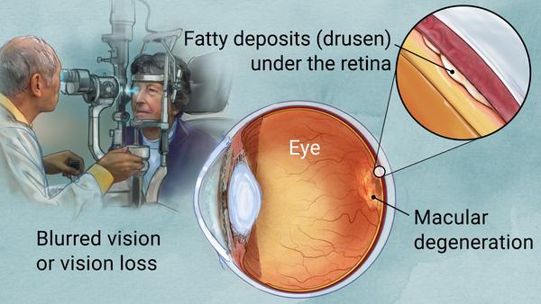
Have an eye check within Omole Akiode Ojodu Berger for detection of Age Related Macular Degeneration (ARMD)

Age-related macular degeneration (AMD) is the most common cause of irreversible central vision loss in older patients. Dilated funduscopic findings are diagnostic; color photographs, fluorescein angiography, and optical coherence tomography assist in confirming the diagnosis and in directing treatment. Treatment is with dietary supplements, intravitreal injection of antivascular endothelial growth factor drugs, laser photocoagulation, photodynamic therapy, and low-vision devices.

Risk factors include the following:

  • Age

  • Genetic variants (eg, abnormal complement factor H)

  • Family history

  • Smoking

  • Obesity

  • Sun exposure

  • A diet low in omega-3 fatty acids and dark green leafy vegetables

    Age-Related Macular Degeneration (AMD or ARMD)

    (Senile Macular Degeneration)

    By

    Sonia Mehta

    , MD, Vitreoretinal Diseases and Surgery Service, Wills Eye Hospital, Sidney Kimmel Medical College at Thomas Jefferson University

    Professional.Manuals.TopicPage.LastRevisionDate| Content last modified Jun 2019
    CLICK HERE FOR PATIENT EDUCATION


    Age-related macular degeneration (AMD) is the most common cause of irreversible central vision loss in older patients. Dilated funduscopic findings are diagnostic; color photographs, fluorescein angiography, and optical coherence tomography assist in confirming the diagnosis and in directing treatment. Treatment is with dietary supplements, intravitreal injection of antivascular endothelial growth factor drugs, laser photocoagulation, photodynamic therapy, and low-vision devices.


    AMD is the leading cause of permanent, irreversible vision loss in older adults. It is more common among whites.


    Two different forms occur:

    • Dry (nonexudative or atrophic): All AMD starts as the dry form. About 85% of people with AMD have only dry AMD.

    • Wet (exudative or neovascular): Wet AMD occurs in about 15% of people.

    Normal Retina
    Normal Retina
    IMAGE PROVIDED BY SUNIR GARG, MD.

Contact us

Eye clinic locations:

Eye centre Akute Ajuwon Fagba Alagbole Agege Alagbado Matogun Olambe Lambe Obawole Giwa Okearo abule Egba Ogba

01, Ajuwon junction, Ajuwon bus stop, beside BPNL filling station, Akute/Ajuwon road, off Elliot bus stop, Iju-Ishagah. Tel: 08034971582

222, Iju road, Balogun, Ifako Ijaye, Lagos. Tel: 08107531046

01, Itshekiri street, beside M. R. S. Petrol station, Ajuwon, Ifo, Ogun state. Tel: 08068084591.

Astigmatism

According to American Optometric Association (AOA), Astigmatism is a common vision condition that causes blurred vision. It occurs when the cornea (the clear front cover of the eye) is irregularly shaped or sometimes because of the curvature of the lens inside the eye.

An irregularly shaped cornea or lens prevents light from focusing properly on the retina, the light-sensitive surface at the back of the eye. As a result, vision becomes blurred at any distance. This can lead to eye discomfort and headaches.

Astigmatism frequently occurs with other vision conditions like myopia (nearsightedness) and hyperopia (farsightedness). Together these vision conditions are referred to as refractive errors because they affect how the eyes bend or “refract” light.

There are many causes to astigmatism. It can be hereditary and is usually present from birth. It can decrease or increase over time.

A comprehensive optometric examination will include testing for astigmatism. If necessary, your optometrist can provide eyeglasses or contact lenses that correct the astigmatism by altering the way light enters the eyes.

Another option for treating astigmatism is a corneal procedure called orthokeratology (ortho-k). In this painless, noninvasive procedure, the patient wears a series of specially designed rigid contact lenses to gradually reshape the curvature of the cornea.

Laser surgery can also treat some types of astigmatism. The laser changes the shape of the cornea by removing a small amount of eye tissue.

[back to top]

What causes astigmatism?

The curvature of the cornea and lens bends the light entering the eye in order to focus it precisely on the retina at the back of the eye. In astigmatism, the surface of the cornea or lens has a somewhat different curvature.

the surface of the cornea is shaped more like a football instead of round like a basketball, the eye is unable to focus light rays to a single point. Vision becomes out of focus at any distance.

In addition, the curvature of the lens inside the eye can change, resulting in an increase or decrease in astigmatism. This change frequently occurs in adulthood and can precede the development of naturally occurring cataracts.

Sometimes astigmatism may develop following an eye injury or eye surgery.

Astigmatism also occurs due to a relatively rare condition called keratoconus in which the cornea becomes progressively thinner and cone-shaped. This results in a large amount of astigmatism, which causes poor vision that cannot be clearly corrected with eyeglasses. People with keratoconus usually need contact lenses for clear vision and eventually may need a corneal transplant.

An optometrist can diagnose an astigmatism through a comprehensive eye examination. Testing for astigmatism measures how the eyes focus light and determines the power of any optical lenses needed to improve vision. This examination may include:

Visual acuity-When you read letters on a distance chart, you are measuring your visual acuity. Visual acuity is given as a fraction (for example, 20/40). The top number is the standardized testing distance (20 feet) and the bottom number is the smallest letter size read. A person with 20/40 visual acuity would have to get within 20 feet to read a letter that should be seen clearly at 40 feet. Normal distance visual acuity is 20/20.

Keratometry/Topography-A keratometer is the primary instrument used to measure the curvature of the cornea. By focusing a circle of light on the cornea and measuring its reflection, it is possible to determine the exact curvature of that area of the cornea’s surface. This measurement is particularly critical in determining the proper fit for contact lenses. A corneal topographer, which is gaining use, generates a contour map of the cornea and provides even more detail of the cornea’s shape.

Refraction-Using an instrument called a phoropter, your optometrist places a series of lenses in front of your eyes and measures how they focus light. This is performed using a handheld lighted instrument called a retinoscope or an automated instrument that evaluates the approximate focusing power of the eye. Based on your responses, the power is then refined to determine the lenses that allow the clearest vision. Despite improved technology, patient input remains integral in determining vision needs.
With the information from these tests, your optometrist can determine if you have astigmatism. Your optometrist will use these findings, combined with those of other tests performed, to determine the power of any lens correction you need to provide clear, comfortable vision. Once testing is complete, your optometrist can discuss treatment options.

[back to top]

How is astigmatism treated?

People with astigmatism have several options to regain clear vision. They include:

Eyeglasses. People with astigmatism primarily choose eyeglasses to improve their vision. The eyeglasses contain a special cylindrical lens prescription that compensates for the astigmatism. This provides additional power in specific parts of the lens.

Generally, a single-vision lens is prescribed to provide clear vision at all distances. However, patients over age 40 who have presbyopia may need a bifocal or progressive addition lens.

Contact lenses. Some people will have better vision with contact lenses rather than eyeglasses. Contact lenses may provide clearer vision and a wider field of view. However, since contact lenses are worn directly on the eyes, they require regular cleaning and care to safeguard eye health.

Standard soft lenses may not be effective in correcting astigmatism. However, special toric soft contact lenses can correct for many types of astigmatism. Because rigid gas-permeable contact lenses maintain their regular shape while on the cornea, they can compensate for the cornea’s irregular shape and improve vision for people with astigmatism.

Orthokeratology. Orthokeratology (ortho-k) involves the fitting of a series of rigid contact lenses to reshape the cornea. The patient wears contact lenses for limited periods, such as overnight, and then removes them. People with moderate astigmatism may be able to temporarily obtain clear vision without lenses for most of their daily activities. Orthokeratology does not permanently improve vision. If patients stop wearing the retainer lenses, their vision may return to its original condition.

Laser and other refractive surgery procedures. Astigmatism can also be corrected by reshaping the cornea through LASIK (laser in situ keratomileusis) or PRK (photorefractive keratectomy). PRK removes tissue from the superficial and inner layers of the cornea. LASIK removes tissue only from the inner layer of the cornea.
If you have an astigmatism, you have a wide range of options to correct your vision problem. In consultation with your optometrist, you can select the treatment that best meets your visual and lifestyle needs

Hyperopia/hypermetropia (longsightedness or farsightedness): How to correct for it

Hyperopia, or farsightedness, is a common vision problem, affecting about a fourth of the population. People with hyperopia can see distant objects very well, but have difficulty focusing on objects that are up close. The condition is sometimes referred to as “hypermetropia” rather than hyperopia.

Hyperopia symptoms

Farsighted people sometimes have headaches or eye strain and may squint or feel fatigued when performing work at close range. If you get these symptoms while wearing your eyeglasses or contact lenses, you may need an eye exam and a new prescription.

What causes hyperopia/hypermetropia

  1. Signs
  2. Causes
  3. Treatment

hyperopia eyeglasses on top of a book

Schedule an exam

FIND EYE DOCTOR

Hyperopia, or farsightedness, is a common vision problem, affecting about a fourth of the population. People with hyperopia can see distant objects very well, but have difficulty focusing on objects that are up close. The condition is sometimes referred to as “hypermetropia” rather than hyperopia.

Hyperopia symptoms

Farsighted people sometimes have headaches or eye strain and may squint or feel fatigued when performing work at close range. If you get these symptoms while wearing your eyeglasses or contact lenses, you may need an eye exam and a new prescription.

What causes hyperopia/hypermetropia?

Watch this video on what causes blurry vision and how we can correct it.

This vision problem occurs when light rays entering the eye focus behind the retina, rather than directly on it. The eyeball of a farsighted person is shorter than normal.

Many children are born farsighted, and some of them “outgrow” it as the eyeball lengthens with normal growth.

Sometimes people confuse hyperopia with presbyopia, which also causes near vision problems but for different reasons.

Hyperopia treatment

Farsightedness can be corrected with glasses or contact lenses to change the way light rays bend into the eyes.

If your glasses or contact lens prescription begins with plus numbers, like +2.50, you are farsighted.

You may need to wear your glasses or contacts all the time or only when reading, working on a computer or doing other close-up work.

When selecting eyeglasses for the correction of farsightedness, choose aspheric high-index lenses — especially for stronger prescriptions. These lenses are thinner, lighter, and have a slimmer, more attractive profile. Aspheric lenses also reduce the magnified “bug-eye” appearance eyeglasses for hyperopia often cause.

Be aware, though, that high-index aspheric lenses reflect more light than standard plastic lenses. For the best comfort and appearance, make sure the lenses include anti-reflective coating, which eliminates distracting lens reflections.

High-index aspheric lenses for children should be made of lightweight polycarbonate lens material for superior comfort and impact resistance.

Also, photochromic lenses that automatically darken in response to sunlight are highly recommended for kids and anyone who spends a significant amount of time outdoors.

Refractive surgery, such as LASIK or CK, is another option for correcting hyperopia. Surgery may reduce or eliminate your need to wear glasses or contact lenses.

Investigational procedures involving corneal implants may be a future option for correcting hyperopia.