Eye clinics near Alagbado Jankara Giwa Okearo Ijoko road Ojokoro

Sex and the eyes: HIV/AIDS and the Eyes

The human immunodeficiency virus (HIV) can cause hemorrhages and strokes in the retina. Having HIV or AIDS can also open the door for a more serious retinal infection from a virus called cytomegalovirus (CMV). CMV infection can lead to retinal detachment and blindness. AIDS can also cause a cancer called Kaposi’s sarcoma that can appear on the eyelids or conjunctiva. HIV infection can also make it easier for a person to get serious infections from other STDs.

Condoms and safer sex can reduce your risk of contracting HIV. Once a person has HIV or AIDS it can affect their eyes by traveling through the blood and body tissue.


If you have any eye infection or discomfort with the eyes, call or visit:

Eye update Eye clinic & opticals01, Ajuwon junction, Ajuwon bus stop, beside BNPL filling station, Akute/Ajuwon road, off Elliot bus stop, Iju-Ishagah near Alagbole Fagba. Tel: 0803497158, 08107531046.

This information will reach people within the following areas:

Obawole, fagba, ifako,ijaye, near, ancillia,agege, iju, road,alagbole, akute, ajuwon, lambe, matogun, matogbun, giwa, okearo, koye, oke, arifanla, baale, Road, fagba, abule, egba, agege, ogba, ikeja, G. R. A, oshodi, Berger, ojodu, omole, magodo, yaba, mowe, ibafo, shagamu, Ishagah, shomolu, heritage, estate, shagamu, bola ahmed tinubu road, kudirat abiola estate, grailand estate, heritage estate, ajao, College Road, iju, Road, water, works, omole, magado, ketu, ojota, ikoyi, akiode bus stop, obawale, grammar school, first gate, Ala Hausa, Yoruba, Igbo, Benin, Edo, Association, Club, Ikorodu, Ketu, Ojota, Elliot, Iju, Ishagah, Fagba, ojodu, best, eye clinic, optical, toll gate, Ikeja, ojota, maryland, oshodi, apapa, ogba, College Road, near bank, station, agege, Abeokuta, sango ota, otta, Lisa, mowe ibafo, redeem camp, Lagos, Ibadan, gold valley estate, isheri, iseri pole phase 1, implemented phase II, grammar school, testing ground, jankara, alagbado, koye oke, oshere, osere, matogun, matogbun, olambe, lambe, tipper garage, otun, jolasco, Victoria Island, festac, vgc, eko atlantic, Victoria garden City, ikorodu, ketu, ajegunle, abule egba, fagba, grailand, Berger, Obawale,Agege, Pencinema,Shomolu, Cele, Ogun, Lagos, Lagos Island, Lagos mainland, Ifako, Ijaye, eti osa, Oyo, Lisa, Owode, sang ota, abule egba, redeem camp, Ijebu ode, toll gate, bus stop, Street, along, Nigeria

Arifanla Akute -Eye clinic near Arifanla Akute Ogun state

Greetings from Eyeupdate Eye clinic & optical supplies.

This page is all about eye care services for Arifanla residents in Akute.

If you have any issues with your eyes, worry no more. Simply contact us at Eyeupdate Eye clinic by filling the form below. You can also call us via 08034971582 or 08107531046 for eyecare advice. Our well equipt eye clinic is located at 01 Ajuwon junction, Ajuwon bus stop near Grail-land Estate gate, Ajuwon

When to see an Eye-Doctor

1, You always have a headache
2, Your eye is infected
3, There are bright flashes or floaters
4, There is eye pain
5, You have been squinting
6, You have diabetes or it is in the family
7, You have hypertension or it is in the family
8, Excessive light sensitivity
9, Your eyes are always red
10, You spend so much time on computer or phone-screen
11, If you have never had your eyes examined before.

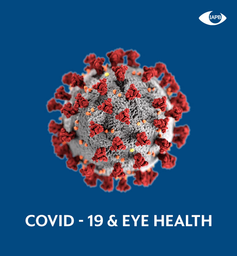
COVID-19 & Eye Health -Eye/Optical within Obawole Iju Ishaga

The 2019 novel coronavirus (COVID-19) pandemic is rapidly changing the way the health and development communities are working. With the proximity of eye health professionals to patients during eye examinations and reports that the virus can cause conjunctivitis, COVID-19 has implications for eye health and eye health professionals. Travel restrictions, reprioritisation of health resources and economic consequences are impacting the work of many of our member and partner organisations.

To assist IAPB members, eye health personnel, health professionals and program personnel, IAPB is collating and sharing information and resources specific to eye health and international development in relation to COVID-19. Without much ado, COVID-19 Resources: Here is what we know:

COVID 19 & Eye Health Resources
COVID 19 Resources
RESOURCES 

Eye Health, Health, International Development and Vision Impairment News and Blogs

Member Stories
MEMBER STORIES

Read the COVID 19 stories from our Members across the globe.

Computer vision syndrome -Eye/Optical clinic near Ojodu Berger Obawole Omole

Computer Vision Syndrome, also referred to as Digital Eye Strain, describes a group of eye and vision-related problems that result from prolonged computer, tablet, e-reader and cell phone use. Many individuals experience eye discomfort and vision problems when viewing digital screens for extended periods. The level of discomfort appears to increase with the amount of digital screen use.

The average American worker spends seven hours a day on the computer either in the office or working from home. March is Save Your Vision Month and the American Optometric Association is working to educate both employers and employees about how to avoid digital eye strain in the workplace. To help alleviate digital eye strain, follow the 20-20-20 rule; take a 20-second break to view something 20 feet away every 20 minutes.

Click here for helpful infographics about the 20-20-20 rule and digital eye strain.

Woman experiencing eye strainThe most common symptoms associated with Computer Vision Syndrome (CVS) or Digital Eye Strain are

  • eyestrain
  • headaches
  • blurred vision
  • dry eyes
  • neck and shoulder pain

These symptoms may be caused by:

  • poor lighting
  • glare on a digital screen
  • improper viewing distances
  • poor seating posture
  • uncorrected vision problems
  • a combination of these factors
  1. The extent to which individuals experience visual symptoms often depends on the level of their visual abilities and the amount of time spent looking at a digital screen. Uncorrected vision problems like farsightedness and astigmatism, inadequate eye focusing or eye coordination abilities, and aging changes of the eyes, such as presbyopia, can all contribute to the development of visual symptoms when using a computer or digital screen device.Many of the visual symptoms experienced by users are only temporary and will decline after stopping computer work or use of the digital device. However, some individuals may experience continued reduced visual abilities, such as blurred distance vision, even after stopping work at a computer. If nothing is done to address the cause of the problem, the symptoms will continue to recur and perhaps worsen with future digital screen use.Prevention or reduction of the vision problems associated with Computer Vision Syndrome or Digital Eye Strain involves taking steps to control lighting and glare on the device screen, establishing proper working distances and posture for screen viewing, and assuring that even minor vision problems are properly corrected.What causes Computer Vision Syndrome or Digital Eye Strain?Viewing a computer or digital screen often makes the eyes work harder. As a result, the unique characteristics and high visual demands of computer and digital screen device viewing make many individuals susceptible to the development of vision-related symptoms.Children using digital devicesUncorrected vision problems can increase the severity of Computer Vision Syndrome or Digital Eye Strain symptoms.Viewing a computer or digital screen is different than reading a printed page. Often the letters on the computer or handheld device are not as precise or sharply defined, the level of contrast of the letters to the background is reduced, and the presence of glare and reflections on the screen may make viewing difficult.

    Viewing distances and angles used for this type of work are also often different from those commonly used for other reading or writing tasks. As a result, the eye focusing and eye movement requirements for digital screen viewing can place additional demands on the visual system.

    In addition, the presence of even minor vision problems can often significantly affect comfort and performance at a computer or while using other digital screen devices. Uncorrected or under corrected vision problems can be major contributing factors to computer-related eyestrain.

    Even people who have an eyeglass or contact lens prescription may find it’s not suitable for the specific viewing distances of their computer screen. Some people tilt their heads at odd angles because their glasses aren’t designed for looking at a computer. Or they bend toward the screen in order to see it clearly. Their postures can result in muscle spasms or pain in the neck, shoulder or back.

    In most cases, symptoms of CVS or Digital Eye Strain occur because the visual demands of the task exceed the visual abilities of the individual to comfortably perform them. At greatest risk for developing CVS or Digital Eye Strain are those persons who spend two or more continuous hours at a computer or using a digital screen device every day.

    How is Computer Vision Syndrome or Digital Eye Strain diagnosed?

    Computer Vision Syndrome, or Digital Eye Strain, can be diagnosed through a comprehensive eye examination. Testing, with special emphasis on visual requirements at the computer or digital device working distance, may include:

  • Patient history to determine any symptoms the patient is experiencing and the presence of any general health problems, medications taken, or environmental factors that may be contributing to the symptoms related to computer use.
  • Visual acuity measurements to assess the extent to which vision may be affected.
  • refraction to determine the appropriate lens power needed to compensate for any refractive errors (nearsightedness, farsightedness or astigmatism).
  • Testing how the eyes focus, move and work together. In order to obtain a clear, single image of what is being viewed, the eyes must effectively change focus, move and work in unison. This testing will look for problems that keep your eyes from focusing effectively or make it difficult to use both eyes together.

This testing may be done without the use of eye drops to determine how the eyes respond under normal seeing conditions. In some cases, such as when some of the eyes’ focusing power may be hidden, eye drops may be used. They temporarily keep the eyes from changing focus while testing is done.

Using the information obtained from these tests, along with results of other tests, your optometrist can determine if you have Computer Vision Syndrome or Digital Eye Strain and advise you on treatment options.

How is Computer Vision Syndrome, or Digital Eye Strain treated?

Solutions to digital screen-related vision problems are varied. However, they can usually be alleviated by obtaining regular eye care and making changes in how you view the screen.

Eye Care

In some cases, individuals who do not require the use of eyeglasses for other daily activities may benefit from glasses prescribed specifically for computer use. In addition, persons already wearing glasses may find their current prescription does not provide optimal vision for viewing a computer.

  • Eyeglasses or contact lenses prescribed for general use may not be adequate for computer work. Lenses prescribed to meet the unique visual demands of computer viewing may be needed. Special lens designs, lens powers or lens tints or coatings may help to maximize visual abilities and comfort.
  • Some computer users experience problems with eye focusing or eye coordination that can’t be adequately corrected with eyeglasses or contact lenses. A program of vision therapy may be needed to treat these specific problems. Vision therapy, also called visual training, is a structured program of visual activities prescribed to improve visual abilities. It trains the eyes and brain to work together more effectively. These eye exercises help remediate deficiencies in eye movement, eye focusing and eye teaming and reinforce the eye-brain connection. Treatment may include office-based as well as home training procedures.


Viewing the Computer

Computer vision syndrome
Proper body positioning for computer use.

Some important factors in preventing or reducing the symptoms of CVS have to do with the computer and how it is used. This includes lighting conditions, chair comfort, location of reference materials, position of the monitor, and the use of rest breaks.

  • Location of computer screen – Most people find it more comfortable to view a computer when the eyes are looking downward. Optimally, the computer screen should be 15 to 20 degrees below eye level (about 4 or 5 inches) as measured from the center of the screen and 20 to 28 inches from the eyes.
  • Reference materials – These materials should be located above the keyboard and below the monitor. If this is not possible, a document holder can be used beside the monitor. The goal is to position the documents so you do not need to move your head to look from the document to the screen.
  • Lighting – Position the computer screen to avoid glare, particularly from overhead lighting or windows. Use blinds or drapes on windows and replace the light bulbs in desk lamps with bulbs of lower wattage.
  • Anti-glare screens – If there is no way to minimize glare from light sources, consider using a screen glare filter. These filters decrease the amount of light reflected from the screen.
  • Seating position – Chairs should be comfortably padded and conform to the body. Chair height should be adjusted so your feet rest flat on the floor. If your chair has arms, they should be adjusted to provide arm support while you are typing. Your wrists shouldn’t rest on the keyboard when typing.
  • Rest breaks – To prevent eyestrain, try to rest your eyes when using the computer for long periods. Rest your eyes for 15 minutes after two hours of continuous computer use. Also, for every 20 minutes of computer viewing, look into the distance for 20 seconds to allow your eyes a chance to refocus.
  • Blinking – To minimize your chances of developing dry eye when using a computer, make an effort to blink frequently. Blinking keeps the front surface of your eye moist.

Regular eye examinations and proper viewing habits can help to prevent or reduce the development of the symptoms associated with Computer Vision Syndrome.


If you are having signs or symptoms of computer vision syndrome, call or visit:

Eye update Eye clinic & opticals 01, Ajuwon junction, Ajuwon bus stop, beside BNPL filling station, Akute/Ajuwon road, off Elliot bus stop, Iju-Ishagah near Alagbole Fagba. Tel: 0803497158, 08107531046.

This information will reach people within the following areas and yonder:

Obawole, fagba, ifako,ijaye, near, ancillia,agege, iju, road,alagbole, akute, ajuwon, lambe, matogun, matogbun, giwa, okearo, koye, oke, arifanla, baale, Road, fagba, abule, egba, agege, ogba, ikeja, G. R. A, oshodi, Berger, ojodu, omole, magodo, yaba, mowe, ibafo, shagamu, Ishagah, shomolu, heritage, estate, shagamu, bola ahmed tinubu road, kudirat abiola estate, grailand estate, heritage estate, ajao, College Road, iju, Road, water, works, omole, magado, ketu, ojota, ikoyi, akiode bus stop, obawale, grammar school, first gate, Ala Hausa, Yoruba, Igbo, Benin, Edo, Association, Club, Ikorodu, Ketu, Ojota, Elliot, Iju, Ishagah, Fagba, ojodu, best, eye clinic, optical, toll gate, Ikeja, ojota, maryland, oshodi, apapa, ogba, College Road, near bank, station, agege, Abeokuta, sango ota, otta, Lisa, mowe ibafo, redeem camp, Lagos, Ibadan, gold valley estate, isheri, iseri pole phase 1, implemented phase II, grammar school, testing ground, jankara, alagbado, koye oke, oshere, osere, matogun, matogbun, olambe, lambe, tipper garage, otun, jolasco, Victoria Island, festac, vgc, eko atlantic, Victoria garden City, ikorodu, ketu, ajegunle, abule egba, fagba, grailand, Berger, Obawale,Agege, Pencinema,Shomolu, Cele, Ogun, Lagos, Lagos Island, Lagos mainland, Ifako, Ijaye, eti osa, Oyo, Lisa, Owode, sang ota, abule egba, redeem camp, Ijebu ode, toll gate, bus stop, Street, along, Nigeria

Contact lenses -Eye/Optical clinic near Obawole Fagba Iju Ishaga

Contact lenses are an excellent choice for nearly anyone who needs vision correction and doesn’t want to wear eyeglasses full time or undergo LASIK surgery.

Unsure about contact lenses? This article will detail contact lens materials, contact lens designs and features and even new contact lens formats.

For instance, the first light-adaptive contact lenses, Acuvue Oasys with Transitions, debuted in the United States in 2019 and contact lenses embedded with antibiotics are in the works. (See our contact lens news page for the latest in contact lenses.)

Here are the basics you should know about contact lenses before seeing your eye doctor if you are interested in wearing contacts.

Contact Lens Materials

The first choice when considering contact lenses is which lens material will best satisfy your needs. There are five types of contact lenses, based on type of lens material they are made of:

ExpandableContact Lens Material

  • Soft lenses are made from gel-like, water-containing plastics called hydrogels. These lenses are very thin and pliable and conform to the front surface of the eye. Introduced in the early 1970s, hydrogel lenses made contact lens wear much more popular because they typically are immediately comfortable. The only alternative at the time was hard contact lenses made of PMMA plastic (see below). PMMA lenses typically took weeks to adapt to and many people couldn’t wear them successfully.
  • Silicone hydrogel lenses are an advanced type of soft contact lenses that are more porous than regular hydrogel lenses and allow even more oxygen to reach the cornea. Introduced in 2002, silicone hydrogel contact lenses are now the most popular lenses prescribed in the United States.
  • Gas permeable lenses — also called GP or RGP lenses — are rigid contact lenses that look and feel like PMMA lenses (see below) but are porous and allow oxygen to pass through them. Because they are permeable to oxygen, GP lenses can be fit closer to the eye than PMMA lenses, making them more comfortable than conventional hard lenses. Since their introduction in 1978, gas permeable contact lenses have essentially replaced nonporous PMMA contact lenses. GP contacts often provide sharper vision than soft and silicone hydrogel contacts — especially if you have astigmatism. It usually takes some time for your eyes to adjust to gas permeable lenses when you first start wearing them, but after this initial adaptation period, most people find GP lenses are as comfortable as hydrogel lenses.
  • Hybrid contact lenses are designed to provide wearing comfort that rivals soft or silicone hydrogel lenses, combined with the crystal-clear optics of gas permeable lenses. Hybrid lenses have a rigid gas permeable central zone, surrounded by a “skirt” of hydrogel or silicone hydrogel material. Despite these features, only a small percentage of people in the U.S. wear hybrid contact lenses, perhaps because these lenses are more difficult to fit and are more expensive to replace than soft and silicone hydrogel lenses.
  • PMMA lenses are made from a transparent rigid plastic material called polymethyl methacrylate (PMMA), which also is used as a substitute for glass in shatterproof windows and is sold under the trademarks Lucite, Perspex and Plexiglas. PMMA lenses have excellent optics, but they do not transmit oxygen to the eye and can be difficult to adapt to. These (now old-fashioned) “hard contacts” have virtually been replaced by GP lenses and are rarely prescribed today.

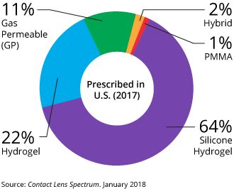
In 2017, 64 percent of contact lenses prescribed in the U.S. were silicone hydrogel lenses, followed by soft (hydrogel) lenses (22 percent), gas permeable lenses (11 percent), hybrid lenses (2 percent) and PMMA lenses (1 percent).


Home Contact Lenses Soft Contact Lenses |  En Español

Contact lens basics: Types of contact lenses and more

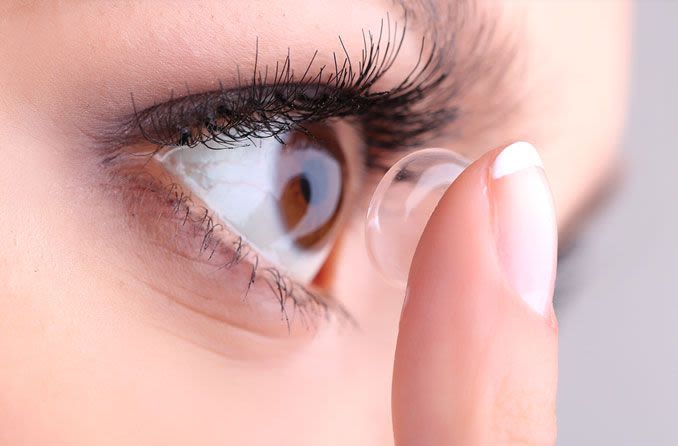
woman applying a contact lens

Contact lenses are an excellent choice for nearly anyone who needs vision correction and doesn’t want to wear eyeglasses full time or undergo LASIK surgery.

Unsure about contact lenses? This article will detail contact lens materials, contact lens designs and features and even new contact lens formats.

For instance, the first light-adaptive contact lenses, Acuvue Oasys with Transitions, debuted in the United States in 2019 and contact lenses embedded with antibiotics are in the works. (See our contact lens news page for the latest in contact lenses.)

Here are the basics you should know about contact lenses before seeing your eye doctor if you are interested in wearing contacts.

Contact Lens Materials

The first choice when considering contact lenses is which lens material will best satisfy your needs. There are five types of contact lenses, based on type of lens material they are made of:

ExpandableContact Lens Material

  • Soft lenses are made from gel-like, water-containing plastics called hydrogels. These lenses are very thin and pliable and conform to the front surface of the eye. Introduced in the early 1970s, hydrogel lenses made contact lens wear much more popular because they typically are immediately comfortable. The only alternative at the time was hard contact lenses made of PMMA plastic (see below). PMMA lenses typically took weeks to adapt to and many people couldn’t wear them successfully.
  • Silicone hydrogel lenses are an advanced type of soft contact lenses that are more porous than regular hydrogel lenses and allow even more oxygen to reach the cornea. Introduced in 2002, silicone hydrogel contact lenses are now the most popular lenses prescribed in the United States.
  • Gas permeable lenses — also called GP or RGP lenses — are rigid contact lenses that look and feel like PMMA lenses (see below) but are porous and allow oxygen to pass through them. Because they are permeable to oxygen, GP lenses can be fit closer to the eye than PMMA lenses, making them more comfortable than conventional hard lenses. Since their introduction in 1978, gas permeable contact lenses have essentially replaced nonporous PMMA contact lenses. GP contacts often provide sharper vision than soft and silicone hydrogel contacts — especially if you have astigmatism. It usually takes some time for your eyes to adjust to gas permeable lenses when you first start wearing them, but after this initial adaptation period, most people find GP lenses are as comfortable as hydrogel lenses.
  • Hybrid contact lenses are designed to provide wearing comfort that rivals soft or silicone hydrogel lenses, combined with the crystal-clear optics of gas permeable lenses. Hybrid lenses have a rigid gas permeable central zone, surrounded by a “skirt” of hydrogel or silicone hydrogel material. Despite these features, only a small percentage of people in the U.S. wear hybrid contact lenses, perhaps because these lenses are more difficult to fit and are more expensive to replace than soft and silicone hydrogel lenses.
  • PMMA lenses are made from a transparent rigid plastic material called polymethyl methacrylate (PMMA), which also is used as a substitute for glass in shatterproof windows and is sold under the trademarks Lucite, Perspex and Plexiglas. PMMA lenses have excellent optics, but they do not transmit oxygen to the eye and can be difficult to adapt to. These (now old-fashioned) “hard contacts” have virtually been replaced by GP lenses and are rarely prescribed today.

In 2017, 64 percent of contact lenses prescribed in the U.S. were silicone hydrogel lenses, followed by soft (hydrogel) lenses (22 percent), gas permeable lenses (11 percent), hybrid lenses (2 percent) and PMMA lenses (1 percent).

LEARN MORE about the proper care of your contact lenses.

Contact Lens Wearing Time

Until 1979, everyone who wore contact lenses removed and cleaned them nightly. The introduction of “extended wear” enabled wearers to sleep in their contacts. Now, two types of lenses are classified by wearing time:

  • Daily wear — must be removed nightly
  • Extended wear — can be worn overnight, usually for seven days consecutively without removal

“Continuous wear” is a term that’s sometimes used to describe 30 consecutive nights of lens wear — the maximum wearing time approved by the FDA for certain brands of extended wear lenses

When To Replace Your Contact Lenses

Even with proper care, contact lenses (especially soft contacts) should be replaced frequently to prevent the build-up of lens deposits and contamination that increase the risk of eye infections.

Soft lenses have these general classifications, based on how frequently they should be discarded:

ExpandableLens Replacement Frequency

  • Daily disposable lenses — Discard after a single day of wear
  • Disposable lenses — Discard every two weeks, or sooner
  • Frequent replacement lenses — Discard monthly or quarterly
  • Traditional (reusable) lenses — Discard every six months or longer

Gas permeable contact lenses are more resistant to lens deposits and don’t need to be discarded as frequently as soft lenses. Often, GP lenses can last a year or longer before they need to be replaced.

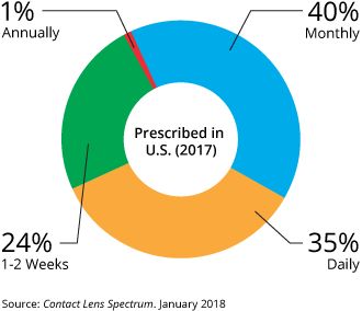
The most frequently prescribed contact lens replacement schedule in the U.S. in 2017 was monthly (40 percent), followed by daily (35 percent), every one to two weeks (24 percent) and annually (1 percent).

LEARN MORE about the proper care of your contact lenses.

Contact Lens Designs

Soft contact lenses (both standard hydrogel and silicone hydrogel lenses) are available in a variety of designs, depending on their intended purpose:

ExpandableSoft contact lens designs

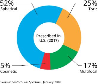
  • Spherical contact lenses have the same lens power throughout the entire optical part of the lens to correct myopia (nearsightedness) or hyperopia (farsightedness).
  • Toric soft contact lenses have different powers in different meridians of the lens to correct astigmatism as well as nearsightedness or farsightedness. [Read more about toric contact lenses.]
  • Multifocal contact lenses (including bifocal contacts) contain different power zones for near and far vision to correct presbyopia as well as nearsightedness or farsightedness. Some multifocal lenses also can correct astigmatism. [Read more about bifocal contacts.]
  • Cosmetic contact lenses include color contacts designed to change or intensify your eye color. Halloween, theatrical and other special-effect contacts also are considered cosmetic lenses. A contact lens prescription is required for cosmetic contacts even if you have no refractive errors that need correction.

All of these lenses can be custom made for hard-to-fit eyes. Other lens designs also are available — including lenses fabricated for use in special situations, such as correcting for keratoconus.

More Contact Lens Features

Bifocal contacts for astigmatism. These are advanced soft contacts that correct both presbyopia and astigmatism, so you can remain glasses-free after age 40 even if you have astigmatism. [More about bifocal contact lenses for astigmatism.]

Contacts for dry eyes. Are your contacts uncomfortably dry? Certain soft contact lenses are specially made to reduce the risk of contact lens-related dry eye symptoms. [More about contact lenses for dry eyes.]

Colored lenses. Many of the types of lenses described above also come in colors that can enhance the natural color of your eyes — that is, make your green eyes even greener, for example. Other colored lenses can totally change the color of your eyes, as in from brown to blue.

Special-effect lenses. Also called theatrical, novelty, or costume lenses, special-effect contacts take coloration one step further to make you look like a cat, a vampire, or another alter-ego of your choice.

Prosthetic lenses. Colored contact lenses also can be used for more medically oriented purposes. Opaque soft lenses called prosthetic contacts can be custom-designed for an eye that has been disfigured by injury or disease to mask the disfigurement and match the appearance of the other, unaffected eye.

Custom lenses. If conventional contact lenses don’t seem to work for you, you might be a candidate for custom contact lenses that are made-to-order for your individual eye shape and visual needs.

UV-inhibiting lenses. Some soft contact lenses help protect your eyes from the sun’s ultraviolet rays that can cause cataracts and other eye problems. But because contacts don’t cover your entire eye, you still should wear UV-blocking sunglasses outdoors for the best protection from the sun.

Scleral lenses. Large-diameter gas permeable lenses called scleral contacts are specially designed to treat keratoconus and other corneal irregularities, as well as presbyopia.

Myopia control contacts. Special contact lenses are being developed to slow or stop the progression of nearsightedness in children. [More about myopia control.]

Which Contact Lens Is Right for You?

First, your contacts must address the problem that is prompting you to wear lenses in the first place. Your contact lenses must provide good vision by correcting your myopiahyperopiaastigmatism, or some combination of those vision problems.

Second, the lens must fit your eye. To do that, lenses come in tens of thousands of combinations of diameter and curvature. Of course, not every lens brand comes in every “size.”

Your ECP is skilled in evaluating your eye’s physiology, and your eyesight, to determine which lens best satisfies the two criteria above.

Third, you may have another medical need that drives the choice of lens. For example, your ECP might pick a particular lens if your eyes tend to be dry.

Finally, consider your “wish list” of contact lens features — colors, for example, or overnight wear.

When you and your ECP decide on the right lens for you, you’ll be given a contact lens prescription. You’ll be able to buy a supply of lenses from your ECP or from the many other outlets that sell contact lenses. [For more on this, please read “Where’s the Best Place to Buy Contact Lenses?”]

Contact Lens Wear and Care

Caring for your contact lenses — cleaning, disinfecting and storing them — is much easier than it used to be.

A few years ago, you would have needed several bottles of cleaning products, and perhaps enzyme tablets, for proper care. Today, most people can use “multipurpose” solutions — meaning that one product both cleans and disinfects, and is used for storage.

People who are sensitive to the preservatives in multipurpose solutions might need preservative-free systems, such as those containing hydrogen peroxide. These do an excellent job of cleaning contacts, but it’s very important to follow the directions for using them. The solution should not come into contact with your eyes until soaking is complete and the solution is neutralized.

Of course, you can avoid lens care altogether by wearing daily disposable contact lenses.

Contact Lens Problems

Trial and error often is involved in finding the perfect lens for you. People react differently to various lens materials and cleaning solutions.

Also, the correct “parameters” of your lens — that is, power, diameter, and curvature — can be finalized only after you’ve successfully worn the lens. This is especially true for more complex fits involving extra parameters, such as with bifocals or toric contact lenses for astigmatism.

If you experience discomfort or poor vision when wearing contact lenses, chances are that an adjustment or change of lens can help.

Today, more contact lens choices than ever are available to provide comfort, good vision, and healthy eyes. If your eyes or lenses are uncomfortable or you are not seeing well, remove your lenses and visit your eye care professional to explore available remedies for contact lens discomfort.

Buying Contact Lenses

You can buy replacement contact lenses at many places, and some offer a better value than others. Find out more about where to buy contacts and buying contact lenses online.

More Information About Contacts

For more information about contacts, visit our contact lens expert at Eyeupdate Eye clinic, 01 Ajuwon junction beside BPNL Filling station, Akute/Ajuwon road, Off Elliot bus stop, Iju-Ishaga, Lagos/Ogun


Contact lenses & solution

Buy prescriptive contact lenses @ N5000 per pair with solution and storage case. Call 08034971582 to place your order

Eye care tips – Eye/Optical clinic near Obawole Fagba Iju Ishaga

Your eyes are an important part of your health. Most people rely on their eyes to see and make sense of the world around them. But some eye diseases can lead to vision loss, so it is important to identify and treat eye diseases as early as possible. You should get your eyes checked as often as your health care provider recommends it, or if you have any new vision problems. And just as it is important to keep your body healthy, you also need to keep your eyes healthy.

Eye Care Tips

There are things you can do to help keep your eyes healthy and make sure you are seeing your best:

  • Eat a healthy, balanced diet. Your diet should include plenty or fruits and vegetables, especially deep yellow and green leafy vegetables. Eating fish high in omega-3 fatty acids, such as salmon, tuna, and halibut can also help your eyes.
  • Maintain a healthy weight. Being overweight or having obesity increases your risk of developing diabetes. Having diabetes puts you at higher risk of getting diabetic retinopathy or glaucoma.
  • Get regular exercise. Exercise may help to prevent or control diabetes, high blood pressure, and high cholesterol. These diseases can lead to some eye or vision problems. So if you exercise regularly, you can lower your risk of getting these eye and vision problems.
  • Wear Transition lenses. Sun exposure can damage your eyes and raise your risk of cataracts and age-related macular degeneration. Protect your eyes by using sunglasses that block out 99 to 100 percent of both UV-A and UV-B radiation.
  • Wear protective eye wear. To prevent eye injuries, you need eye protection when playing certain sports, working in jobs such as factory work and construction, and doing repairs or projects in your home.
  • Avoid smoking. Smoking increases the risk of developing age-related eye diseases such as macular degeneration and cataracts and can damage the optic nerve.
  • Know your family medical history. Some eye diseases are inherited, so it is important to find out whether anyone in your family has had them. This can help you determine if you are at higher risk of developing an eye disease.
  • Know your other risk factors. As you get older, you are at higher risk of developing age-related eye diseases and conditions. It is important to know you risk factors because you may be able to lower your risk by changing some behaviors.
  • If you wear contacts, take steps to prevent eye infections. Wash your hands well before you put in or take out your contact lenses. Also follow the instructions on how to properly clean them, and replace them when needed.
  • Give your eyes a rest. If you spend a lot of time using a computer, you can forget to blink your eyes and your eyes can get tired. To reduce eyestrain, try the 20-20-20 rule: Every 20 minutes, look away about 20 feet in front of you for 20 seconds.

Eye Tests and Exams

Everyone needs to have their eyesight tested to check for vision and eye problems. Children usually have vision screening in school or at their health care provider’s office during a checkup. Adults may also get vision screenings during their checkups. But many adults need more than a vision screening. They need a comprehensive dilated eye exam.

Getting comprehensive dilated eye exams is especially important because some eye diseases may not have warning signs. The exams are the only way to detect these diseases in their early stages, when they are easier to treat.

The exam includes several tests:

  • A visual field test to measure your side (peripheral) vision. A loss of peripheral vision may be a sign of glaucoma.
  • A visual acuity test, where you read an eye chart about 20 feet away, to check on how well you see at various distances
  • Tonometry, which measures your eye’s interior pressure. It helps to detect glaucoma.
  • Dilation, which involves getting eye drops that dilate (widen) your pupils. This allows more light to enter the eye. Your eye care provider examines your eyes using a special magnifying lens. This provides a clear view of important tissues at the back of your eye, including the retina, macula, and optic nerve.

If you have a refractive error and are going to need glasses or contacts, then you will also have a refraction test. When you have this test, you look through a device that has lenses of different strengths to help your eye care professional figure out which lenses will give you the clearest vision.

At what age you should start getting these exams and how often you need them depends on many factors. They include your age, race, and overall health. For example, if you are African American, you are at higher risk of glaucoma and you need to start getting the exams earlier. If you have diabetes, you should get an exam every year. Check with your health care provider about if and when you need these exams

Eye test – Eye clinic within Obawole Iju Ishagah Fagba

Dr. Dr. Steven E.N., Eyeupdate clinic & optical supplies, 01 Ajuwon junction, beside BPNL filling station, Ajuwon, off Elliot bus stop, Iju-Ishagah, Lagos. Tel: 08107531046, 08034971582.


Eye exam/test consists of series of tests that your eye doctor will perform in order to determine your ocular status.

An eye exam involves a series of tests to evaluate your vision and check for eye diseases/vision defects. Your eye doctor may use a variety of instruments, shine bright lights directly at your eyes and request that you look through an array of lenses. Each test during an eye exam evaluates a different aspect of your vision or eye health.

Why it’s done

An eye exam helps detect eye problems at their earliest stage — when they’re most treatable. Regular eye exams give your eye care professional a chance to help you correct or adapt to vision changes and provide you with tips on caring for your eyes.

When to have an eye exam

Several factors may determine how frequently you need an eye exam, including your age, health and risk of developing eye problems. General guidelines are as follows:

Children 3 years and younger

For children under 3, your pediatrician will likely look for the most common eye problems — lazy eye, crossed eyes or misaligned eyes. If there are eye concerns or symptoms, an examination is appropriate at that time regardless of age. Your child could undergo a more comprehensive eye exam between the ages of 3 and 5.

School-age children and adolescents

Have your child’s vision checked before he or she enters first grade. If your child has no symptoms of vision problems and no family history of vision problems, have his or her vision rechecked every one to two years. Otherwise, schedule eye exams based on the advice of your eye doctor.

Adults

In general, if you are healthy and you have no symptoms of vision problems, have your eyes checked on this schedule:

  • Every five to 10 years in your 20s and 30s
  • Every two to four years from 40 to 54
  • Every one to three years from 55 to 64
  • Every one to two years after age 65

Have your eyes checked more often if you:

  • Wear glasses or contact lenses
  • Have a family history of eye disease or loss of vision
  • Have a chronic disease that puts you at greater risk of eye disease, such as diabetes
  • Take medications that have serious eye side effects
  • How you prepare

    There are three different types of eye specialists. Which specialist you choose may be a matter of personal preference or will depend on the nature of your eye problem.

    • Ophthalmologists. Ophthalmologists are medical doctors who provide full eye care, such as giving you a complete eye exam, prescribing corrective lenses, diagnosing and treating complex eye diseases, and performing eye surgery.
    • Optometrists. Optometrists provide many of the same services as ophthalmologists, such as evaluating your vision, prescribing corrective lenses, diagnosing common eye disorders and treating selected eye diseases with drugs. If you have a complex eye problem or need surgery, your doctor can refer you to an ophthalmologist.
    • Opticians. Opticians fill prescriptions for eyeglasses, including assembling, fitting and selling them. Some opticians also sell contact lenses. Opticians do not provide eye health evaluations.

    Bring your prescription eyewear

    If you wear contact lenses or glasses, bring them to your appointment. Your eye doctor will want to make sure your prescription is the best one for you.

    Other precautions

    If your eyes are dilated as a part of your eye exam, you may want to bring sunglasses to wear after your eye exam is complete, as daylight or other bright lights may be uncomfortable or cause blurred vision. Also, consider having someone else drive you home.

    What you can expect

    Before the exam

    If you’re seeing a new eye doctor or if you’re having your first eye exam, expect questions about your vision history. Part of the examination, such as taking your medical history and the initial eye test, may be performed by a clinical assistant or technician.

    Your answers help your eye doctor understand your risk of eye disease and vision problems. Be prepared to give specific information, including:

    • Are you having any eye problems now?
    • Have you had any eye problems in the past?
    • Do you wear glasses or contacts now? If so, are you satisfied with them?
    • What health problems have you had in recent years?
    • Were you born prematurely?
    • Are you taking any medications?
    • Do you have any allergies to medications, food or other substances?
    • Have you ever had eye surgery?
    • Does anyone in your family have eye problems, such as macular degeneration, glaucoma or retinal detachments?
    • Do you or does anyone in your family have diabetes, high blood pressure, heart disease or any other health problems that can affect the whole body?

    During the exam

    An eye exam usually involves these steps:

    • You’ll be asked about your medical history and any vision problems you might be experiencing.
    • Your eye doctor measures your visual acuity to see if you need glasses or contact lenses to improve your vision.
    • You’ll be given a numbing drop in your eyes. Then your eye pressure is measured. To make it easier for your doctor to examine the inside of your eye, he or she will likely dilate your eyes with eyedrops.
    • After waiting for the dilating drops to take effect, your eye doctor checks the health of your eyes, possibly using several lights to evaluate the front of the eye and the inside of each eye.

    Several different tests may be performed during the eye exam. The tests are designed to check your vision and to examine the appearance and function of all parts of your eyes.

    After the exam

    At the end of your eye exam, you and your doctor will discuss the results of all testing, including an assessment of your vision, your risk of eye disease and preventive measures you can take to protect your eyesight.

    Different types of eye exams

    Eye muscle test

    This test evaluates the muscles that control eye movement. Your eye doctor watches your eye movements as you follow a moving object, such as a pen or small light, with your eyes. He or she looks for muscle weakness, poor control or poor coordination.

    Visual acuity test

    This test measures how clearly you see. Your doctor asks you to identify different letters of the alphabet printed on a chart (Snellen chart) or a screen positioned some distance away. The lines of type get smaller as you move down the chart. Each eye is tested separately. Your near vision also may be tested, using a card with letters similar to the distant eye chart. The card is held at reading distance.

    Refraction assessment

    Light waves are bent as they pass through your cornea and lens. If light rays don’t focus perfectly on the back of your eye, you have a refractive error. Having a refractive error may mean you need some form of correction, such as glasses, contact lenses or refractive surgery, to see as clearly as possible.

    Assessment of your refractive error helps your doctor determine a lens prescription that will give you the sharpest, most comfortable vision. The assessment may also determine that you don’t need corrective lenses.

    Your doctor may use a computerized refractor to estimate your prescription for glasses or contact lenses. Or he or she may use a technique called retinoscopy. In this procedure, the doctor shines a light into your eye and measures the refractive error by evaluating the movement of the light reflected by your retina back through your pupil.

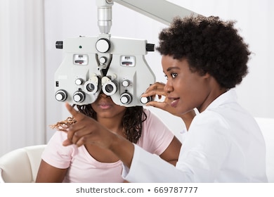
    Your eye doctor usually fine-tunes this refraction assessment by having you look through a masklike device that contains wheels of different lenses (phoropter). He or she asks you to judge which combination of lenses gives you the sharpest vision.

    Visual field test (perimetry)

    Your visual field is the full extent of what you can see to the sides without moving your eyes. The visual field test determines whether you have difficulty seeing in any areas of your overall field of vision. The different types of visual field tests include:

    • Confrontation exam. Your eye doctor sits directly in front of you and asks you to cover one eye. You look straight ahead and tell the doctor each time you see his or her hand move into view.
    • Manual testing, including tangent screen and Goldmann exams. You sit a short distance from a screen and focus on a target at its center. You tell the doctor when you can see an object move into your peripheral vision and when it disappears.
    • Automated perimetry. As you look at a screen with blinking lights on it, you press a button each time you see a blink.

    Using your responses to one or more of these tests, your eye doctor determines the fullness of your field of vision. If you aren’t able to see in certain areas, noting the pattern of your visual field loss may help your eye doctor diagnose your eye condition.

    Color vision testing

    You could have poor color vision and not even realize it. If you have difficulty distinguishing certain colors, your eye doctor may screen your vision for a color deficiency. To do this, your doctor shows you several multicolored dot-pattern tests.

    If you have no color deficiency, you’ll be able to pick out numbers and shapes from within the dot patterns. If you do have a color deficiency, you’ll find it difficult to see certain patterns within the dots. Your doctor may use other tests, as well.

    Slit-lamp examination

    A slit lamp is a microscope that magnifies and illuminates the front of your eye with an intense line of light. Your doctor uses this device to examine the eyelids, lashes, cornea, iris, lens and fluid chamber between your cornea and iris.

    Your doctor may use a dye, most commonly fluorescein (flooh-RES-een), to color the film of tears over your eye. This helps reveal any damaged cells on the front of your eye. Your tears wash the dye from the surface of your eye fairly quickly.

    Retinal examination

    A retinal examination — sometimes called ophthalmoscopy or funduscopy — allows your doctor to evaluate the back of your eye, including the retina, the optic disk and the underlying layer of blood vessels that nourish the retina (choroid). Usually before your doctor can see these structures, your pupils must be dilated with eyedrops that keep the pupil from getting smaller when your doctor shines light into the eye.

    After administering eyedrops and giving them time to work, your eye doctor may use one or more of these techniques to view the back of your eye:

    • Direct exam. Your eye doctor uses an ophthalmoscope to shine a beam of light through your pupil to see the back of the eye. Sometimes eyedrops aren’t necessary to dilate your eyes before this exam.
    • Indirect exam. During this exam, you might lie down, recline in a chair or sit up. Your eye doctor examines the inside of the eye with the aid of a condensing lens and a bright light mounted on his or her forehead. This exam lets your doctor see the retina and other structures inside your eye in great detail and in three dimensions.

    Screening for glaucoma

    Tonometry measures the fluid pressure inside your eye (intraocular pressure). This is one test that helps your eye doctor detect glaucoma, a disease that damages the optic nerve.

    Several methods to measure intraocular pressure are available, including:

    • Applanation tonometry. This test measures the amount of force needed to temporarily flatten a part of your cornea. You’ll be given eyedrops with fluorescein, the same dye used in a regular slit-lamp examination. You’ll also receive eyedrops containing an anesthetic. Using the slit lamp, your doctor moves the tonometer to touch your cornea and determine the eye pressure. Because your eye is numbed, the test doesn’t hurt.
    • Noncontact tonometry. This method uses a puff of air to estimate the pressure in your eye. No instruments touch your eye, so you won’t need an anesthetic. You’ll feel a momentary pulse of air on your eye, which can be startling.

    If your eye pressure is higher than average or your optic nerve looks unusual, your doctor may use a pachometer. This instrument uses sound waves to measure the thickness of your cornea. The most common way of measuring corneal thickness is to put an anesthetic drop in your eye, then place a small probe in contact with the front surface of the eye. The measurement takes seconds.

    You may need more-specialized tests, depending on your age, medical history and risk of developing eye disease.

    Results

    Normal results from an eye exam include:

    • 20/20 vision
    • Good peripheral vision
    • Ability to distinguish various colors
    • Normal-appearing structures of the external eye
    • Absence of cataract, glaucoma or retinal disorders, such as macular degeneration

    Your doctor may give you a prescription for corrective lenses. If your eye exam yields other abnormal results, your doctor will discuss with you next steps for further testing or for treating an underlying condition.

Diabetic retinopathy – Eye clinic near Fagba Obawale Iju-Ishagah

Overview

Diabetic retinopathy (die-uh-BET-ik ret-ih-NOP-uh-thee) is a diabetes complication that affects eyes. It’s caused by damage to the blood vessels of the light-sensitive tissue at the back of the eye (retina).

At first, diabetic retinopathy may cause no symptoms or only mild vision problems. Eventually, it can cause blindness.

The condition can develop in anyone who has type 1 or type 2 diabetes. The longer you have diabetes and the less controlled your blood sugar is, the more likely you are to develop this eye complication.

Symptoms

You might not have symptoms in the early stages of diabetic retinopathy. As the condition progresses, diabetic retinopathy symptoms may include:

  • Spots or dark strings floating in your vision (floaters)
  • Blurred vision
  • Fluctuating vision
  • Impaired color vision
  • Dark or empty areas in your vision
  • Vision loss

Diabetic retinopathy usually affects both eyes.

When to see a doctor

Careful management of your diabetes is the best way to prevent vision loss. If you have diabetes, see your eye doctor for a yearly eye exam with dilation — even if your vision seems fine. Pregnancy may worsen diabetic retinopathy, so if you’re pregnant, your eye doctor may recommend additional eye exams throughout your pregnancy.

Contact your eye doctor right away if your vision changes suddenly or becomes blurry, spotty or hazy.

Causes

Over time, too much sugar in your blood can lead to the blockage of the tiny blood vessels that nourish the retina, cutting off its blood supply. As a result, the eye attempts to grow new blood vessels. But these new blood vessels don’t develop properly and can leak easily.

There are two types of diabetic retinopathy:

  • Early diabetic retinopathy. In this more common form — called nonproliferative diabetic retinopathy (NPDR) — new blood vessels aren’t growing (proliferating).When you have NPDR, the walls of the blood vessels in your retina weaken. Tiny bulges (microaneurysms) protrude from the vessel walls of the smaller vessels, sometimes leaking fluid and blood into the retina. Larger retinal vessels can begin to dilate and become irregular in diameter, as well. NPDR can progress from mild to severe, as more blood vessels become blocked.Nerve fibers in the retina may begin to swell. Sometimes the central part of the retina (macula) begins to swell (macular edema), a condition that requires treatment.
  • Advanced diabetic retinopathy. Diabetic retinopathy can progress to this more severe type, known as proliferative diabetic retinopathy. In this type, damaged blood vessels close off, causing the growth of new, abnormal blood vessels in the retina, and can leak into the clear, jelly-like substance that fills the center of your eye (vitreous).Eventually, scar tissue stimulated by the growth of new blood vessels may cause the retina to detach from the back of your eye. If the new blood vessels interfere with the normal flow of fluid out of the eye, pressure may build up in the eyeball. This can damage the nerve that carries images from your eye to your brain (optic nerve), resulting in glaucoma.

Risk factors

Anyone who has diabetes can develop diabetic retinopathy. Risk of developing the eye condition can increase as a result of:

  • Duration of diabetes — the longer you have diabetes, the greater your risk of developing diabetic retinopathy
  • Poor control of your blood sugar level
  • High blood pressure
  • High cholesterol
  • Pregnancy
  • Tobacco use
  • Being African-American, Hispanic or Native American

Complications

Diabetic retinopathy involves the abnormal growth of blood vessels in the retina. Complications can lead to serious vision problems:

  • Vitreous hemorrhage. The new blood vessels may bleed into the clear, jelly-like substance that fills the center of your eye. If the amount of bleeding is small, you might see only a few dark spots (floaters). In more-severe cases, blood can fill the vitreous cavity and completely block your vision.Vitreous hemorrhage by itself usually doesn’t cause permanent vision loss. The blood often clears from the eye within a few weeks or months. Unless your retina is damaged, your vision may return to its previous clarity.
  • Retinal detachment. The abnormal blood vessels associated with diabetic retinopathy stimulate the growth of scar tissue, which can pull the retina away from the back of the eye. This may cause spots floating in your vision, flashes of light or severe vision loss.
  • Glaucoma. New blood vessels may grow in the front part of your eye and interfere with the normal flow of fluid out of the eye, causing pressure in the eye to build up (glaucoma). This pressure can damage the nerve that carries images from your eye to your brain (optic nerve).
  • Blindness. Eventually, diabetic retinopathy, glaucoma or both can lead to complete vision loss.

Prevention

You can’t always prevent diabetic retinopathy. However, regular eye exams, good control of your blood sugar and blood pressure, and early intervention for vision problems can help prevent severe vision loss.

If you have diabetes, reduce your risk of getting diabetic retinopathy by doing the following:

  • Manage your diabetes. Make healthy eating and physical activity part of your daily routine. Try to get at least 150 minutes of moderate aerobic activity, such as walking, each week. Take oral diabetes medications or insulin as directed.
  • Monitor your blood sugar level. You may need to check and record your blood sugar level several times a day — more-frequent measurements may be required if you’re ill or under stress. Ask your doctor how often you need to test your blood sugar.
  • Ask your doctor about a glycosylated hemoglobin test. The glycosylated hemoglobin test, or hemoglobin A1C test, reflects your average blood sugar level for the two- to three-month period before the test. For most people, the A1C goal is to be under 7 percent.
  • Keep your blood pressure and cholesterol under control. Eating healthy foods, exercising regularly and losing excess weight can help. Sometimes medication is needed, too.
  • If you smoke or use other types of tobacco, ask your doctor to help you quit. Smoking increases your risk of various diabetes complications, including diabetic retinopathy.
  • Pay attention to vision changes. Contact your eye doctor right away if you experience sudden vision changes or your vision becomes blurry, spotty or hazy.

Remember, diabetes doesn’t necessarily lead to vision loss. Taking an active role in diabetes management can go a long way toward preventing complications.

Diagnosis

Diabetic retinopathy is best diagnosed with a comprehensive dilated eye exam. For this exam, drops placed in your eyes widen (dilate) your pupils to allow your doctor to better view inside your eyes. The drops may cause your close vision to blur until they wear off, several hours later.

During the exam, your eye doctor will look for:

  • Abnormal blood vessels
  • Swelling, blood or fatty deposits in the retina
  • Growth of new blood vessels and scar tissue
  • Bleeding in the clear, jelly-like substance that fills the center of the eye (vitreous)
  • Retinal detachment
  • Abnormalities in your optic nerve

In addition, your eye doctor may:

  • Test your vision
  • Measure your eye pressure to test for glaucoma
  • Look for evidence of cataracts

Fluorescein angiography

With your eyes dilated, your doctor takes pictures of the inside of your eyes. Then your doctor will inject a special dye into your arm vein and take more pictures as the dye circulates through your eyes’ blood vessels. Your doctor can use the images to pinpoint blood vessels that are closed, broken down or leaking fluid.

Optical coherence tomography

Your eye doctor may request an optical coherence tomography (OCT) exam. This imaging test provides cross-sectional images of the retina that show the thickness of the retina, which will help determine whether fluid has leaked into retinal tissue. Later, OCT exams can be used to monitor how treatment is working.

Treatment

Treatment, which depends largely on the type of diabetic retinopathy you have and how severe it is, is geared to slowing or stopping progression of the condition.

Early diabetic retinopathy

If you have mild or moderate nonproliferative diabetic retinopathy, you may not need treatment right away. However, your eye doctor will closely monitor your eyes to determine when you might need treatment.

Work with your diabetes doctor (endocrinologist) to determine if there are ways to improve your diabetes management. When diabetic retinopathy is mild or moderate, good blood sugar control can usually slow the progression.

Advanced diabetic retinopathy

If you have proliferative diabetic retinopathy or macular edema, you’ll need prompt surgical treatment. Depending on the specific problems with your retina, options may include:

  • Photocoagulation. This laser treatment, also known as focal laser treatment, can stop or slow the leakage of blood and fluid in the eye. During the procedure, leaks from abnormal blood vessels are treated with laser burns.Focal laser treatment is usually done in your doctor’s office or eye clinic in a single session. If you had blurred vision from macular edema before surgery, the treatment might not return your vision to normal, but it’s likely to reduce the chance the macular edema may worsen.
  • Panretinal photocoagulation. This laser treatment, also known as scatter laser treatment, can shrink the abnormal blood vessels. During the procedure, the areas of the retina away from the macula are treated with scattered laser burns. The burns cause the abnormal new blood vessels to shrink and scar.It’s usually done in your doctor’s office or eye clinic in two or more sessions. Your vision will be blurry for about a day after the procedure. Some loss of peripheral vision or night vision after the procedure is possible.
  • Vitrectomy. This procedure uses a tiny incision in your eye to remove blood from the middle of the eye (vitreous) as well as scar tissue that’s tugging on the retina. It’s done in a surgery center or hospital using local or general anesthesia.
  • Injecting medicine into the eye. Your doctor may suggest injecting medication into the vitreous in the eye. These medications, called vascular endothelial growth factor (VEGF) inhibitors, may help stop growth of new blood vessels by blocking the effects of growth signals the body sends to generate new blood vessels.Your doctor may recommend these medications, also called anti-VEGF therapy, as a stand-alone treatment or in combination with panretinal photocoagulation. While studies of anti-VEGF therapy in the treatment of diabetic retinopathy are promising, this approach is not yet considered standard.Surgery often slows or stops the progression of diabetic retinopathy, but it’s not a cure. Because diabetes is a lifelong condition, future retinal damage and vision loss are still possible.

Even after treatment for diabetic retinopathy, you’ll need regular eye exams. At some point, additional treatment may be recommended.

Clinical trials

Explore Mayo Clinic studies testing new treatments, interventions and tests as a means to prevent, detect, treat or manage this disease.

Alternative medicine

Several alternative therapies have suggested some benefits for people with diabetic retinopathy, but more research is needed to understand whether these treatments are effective and safe.

Be sure to let your doctor know if you are taking any herbs or supplements. They have the potential to interact with other medications, or cause complications in surgery, such as excessive bleeding.

It’s vital not to delay standard treatments to try unproven therapies. Early treatment is the best way to prevent vision loss.

Coping and support

The thought that you might lose your sight can be frightening, and you may benefit from talking to a therapist or finding a support group. Ask your doctor for referrals.

If you’ve already lost vision, ask your doctor about low-vision products, such as magnifiers, and services that can make daily living easier.

Preparing for your appointment

The American Diabetes Association (ADA) recommends that anyone who’s older than 10 with type 1 diabetes have his or her first eye exam within five years of being diagnosed with diabetes.

If you have type 2 diabetes, the ADA advises getting your initial eye exam at the time of your diagnosis, because you may have had diabetes for some time without knowing it.

If there’s no evidence of retinopathy on your initial exam, the ADA recommends that people with diabetes get dilated and comprehensive eye exams at least every two years. If you have any level of retinopathy, you’ll need eye exams at least annually. Ask your eye doctor what he or she recommends.

The ADA recommends that women with diabetes have an eye exam before becoming pregnant or during the first trimester of pregnancy and be closely followed during the pregnancy and up to one year after giving birth. Pregnancy can sometimes cause diabetic retinopathy to develop or worsen.

Here’s some information to help you get ready for your eye appointment.

What you can do

  • Write a brief summary of your diabetes history, including when you were diagnosed; medications you have taken for diabetes, now and in the past; recent average blood sugar levels; and your last few hemoglobin A1C readings, if you know them.
  • List other medications, vitamins and supplements you take, and the dosage.
  • List your symptoms, if any. Include any that may seem unrelated to potential eye problems.
  • Ask a family member or friend to go with you, if possible. Someone who accompanies you can help remember the information you receive. Also, because your eyes have been dilated, a companion can drive you home.
  • List questions for your doctor.

For diabetic retinopathy, some basic questions to ask your doctor include:

  • How is diabetes affecting my vision?
  • Do I need other tests?
  • Is this condition temporary or long lasting?
  • What treatments are available, and which do you recommend?
  • What side effects might I expect from treatment?
  • I have other health conditions. How can I best manage them together?
  • If I control my blood sugar, will my eye symptoms go away?
  • What do my blood sugar goals need to be to protect my eyes?
  • Can you recommend services for people with visual impairment?

Don’t hesitate to ask other questions you have.

What to expect from your doctor

Your doctor is likely to ask you a number of questions, including:

  • Do you have eye symptoms, such as blurred vision or floaters?
  • How long have you had symptoms?
  • In general, how well are you controlling your diabetes?
  • What was your last hemoglobin A1C?
  • Do you have other health conditions, such as high blood pressure or high cholesterol?
  • Have you had eye surgery?

High blood pressure & your eyes – Eye clinic near Sango Ota Abule Egba and Fagba