Two Patients With Macular Degeneration Get Artificial Retinas

LYON, France — A 72-year-old woman with dry age-related macular degeneration (AMD) was the first patient to receive an artificial retina implant as part of the multicenter PRIMAvera clinical trial, which is looking at the safety and efficacy of the PRIMA system.

“Her vision was severely impaired by this condition. We used the ETDRS chart to assess her visual acuity, as this is the current method in ophthalmology. She was able to read only nine letters,” said Laurent Kodjikian, MD, PhD, from Hôpital de la Croix-Rousse in Lyon, who is a former president of the French Society of Ophthalmology. To put that in perspective, a person with normal vision can make out 85 letters, he explained.

“The goal is to get her reading another ten letters,” he told Medscape Medical News.

The hospital where Kodjikian works is one of six centers in France selected to take part in the ongoing AMD clinical trial; other study sites are in Germany, Italy, the Netherlands, and Spain. The 38 study participants will be followed for 12 months after implantation to assess visual acuity and adverse events, and outcomes will be monitored for 3 years. Investigators hope that the findings will lead to the device receiving authorization to enter the market.

The PRIMA artificial retina system has three elements: a tiny wireless retinal implant; a pair of glasses with a camera and digital projector; and a portable processor connected to the projector. The camera captures visual scenes from the surrounding environment. The processor uses algorithms to process and simplify the images, which are then sent back to the glasses. The digital projector uses pulses of infrared light to project the processed images onto the retinal implant’s photovoltaic receptors. These receptors then convert the optical information into electrical stimulation, which excites the nerve cells of the inner retina, allowing them take in the information and transmit it, via the optic nerve, to the brain. This then induces visual perception.

A Delicate Operation

To implant the chip, Kodjikian made a rather large incision — 3.5 mm — and then peeled off the retina, all while looking through a surgical microscope.

“It’s easy to peel off the retina in a healthy eye. However, the procedure becomes more difficult in an eye affected by dry AMD, where the retina tissue is not only very thin and firmly attached to the back wall of the eye, but is also very fragile. A lot can go wrong during this step, so we have to really take our time,” he said. “You can’t go too deep, and if you go too close to the surface, you risk perforating the retina. Like walking on a very thin tightrope, there’s danger all around, and very little room for error.”

After the chip was inserted under the retina, Kodjikian put the retina tissue back into place. “This was the first time I’d ever done this kind of procedure, and it was quite a challenge,” he told Medscape.

The operation took 2.5 hours, which is much less time than the 4 to 5 hours estimated by the manufacturer.

The patient will undergo rehabilitation for 12 months to help her adapt to the system. “Our hope is that this patient will be able to see better with the implant. She probably won’t get to the point of being able to drive a car. And while reading novels in small print may not be possible, it’s quite likely that she’ll be able to read large-print editions,” Kodjikian explained.

Upon activation of the retinal prosthetic, the patient experienced visual impressions that she couldn’t see before the surgery. And 1 month after the procedure, things seem to be on track, according to a press release. “The postoperative result is excellent. There are no complications, the chip is perfectly in place and the vision has not been degraded by the operation. She should now start to improve thanks to rehabilitation,” said Kodjikian.

“Of the various artificial retina systems out there, this one is the most sophisticated because it has the most pixels. The technology will certainly continue to advance. But for the time being, the clinical study should allow us to show that it does work,” he concluded.

In December 2021, he implanted an artificial retina in a second patient, and it took him 50 minutes less than the first one.

This article originally appeared in the French edition of Medscape.

The authors have disclosed no relevant financial relationships.

Eyeupdate Eye-clinic & Optical Supplies, p1, Ajuwon bus stop, Akute/Ajuwon Road, Ajuwon (beside BPNL/MRS Filling station).
Tel: 08107531046, 08034971582.

Our eye-clinic/optical care services cover people from the following locations and near you:
alagbole, akute, ajuwon, lambe, matogun, matogbun, giwa, okearo, koye, oke, arifanla, baale, Road, fagba, abule, egba, agege, ogba, ikeja, G. R. A, oshodi, Berger, ojodu, omole, magodo, yaba, mowe, ibafo, shagamu, Ishagah, shomolu, heritage, estate, ajao, College Road, iju, Road, water, works, omole, magado, ketu, ojota, ikoyi, akiode bus stop, grammar school, first gate, Ala Hausa, Yoruba, Igbo, Benin, Edo, Association, Club, Ikorodu, Ketu, ijoko, jankara, Ladipo, Ojota, Shomolu, Cele, Ogun, Lagos, Lagos Island, Lagos mainland, Ifako, Ijaye, eti osa, Oyo, Lisa, Obawole,Iju, Ishagah, Ishaga, Isaga, Isagah, Ishasi,Obawale,Owode, sang ota, abule egba, redeem camp, Ijebu ode, toll gate, bus stop, Street, along, Nigeria.

Glaucoma

Glaucoma

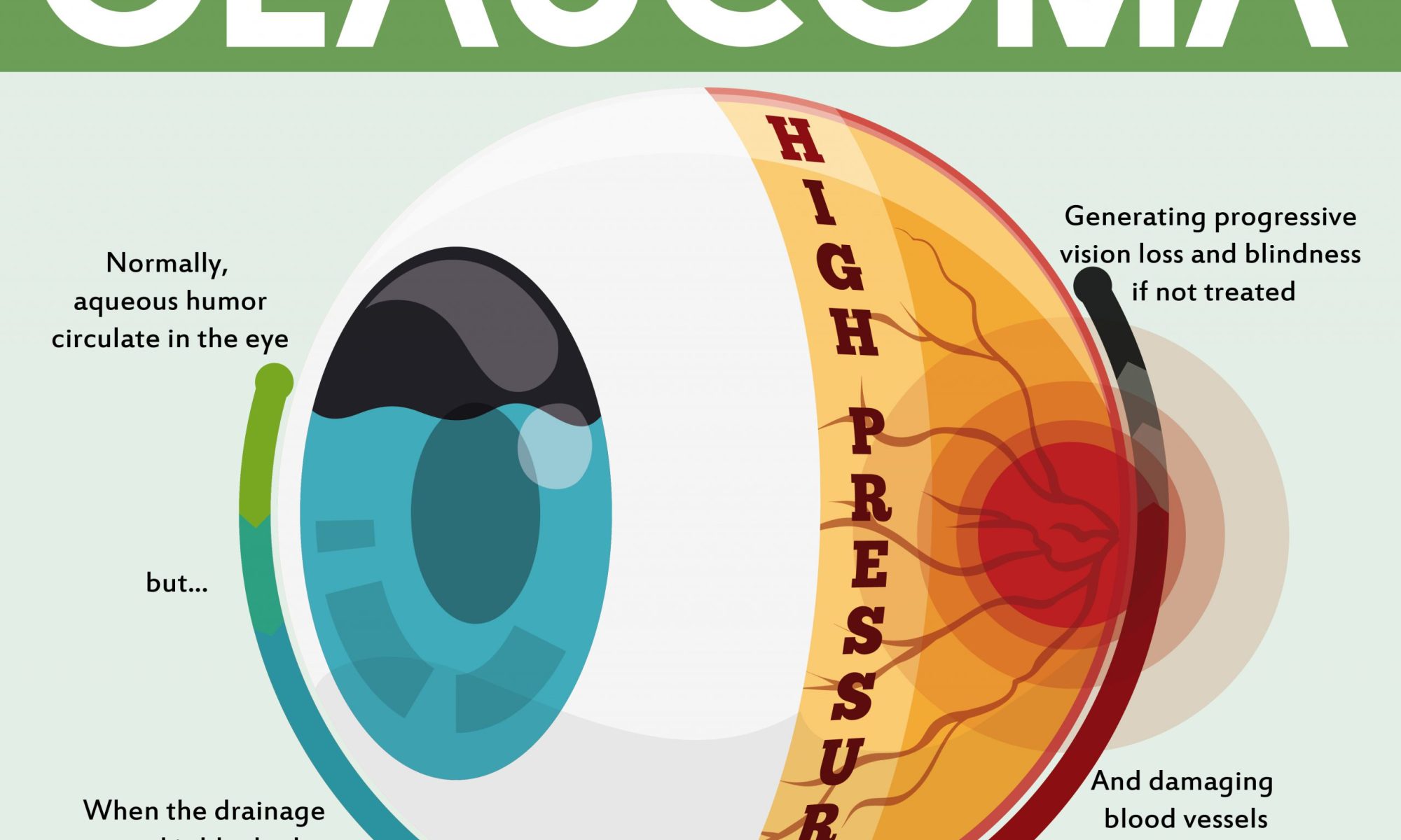
Glaucoma is a condition that damages your eye‘s optic nerve. It gets worse over time. It’s often linked to a buildup of pressure inside your eyeGlaucoma tends to run in families. You usually don’t get it until later in life. 

The increased pressure in your eye, called intraocular pressure, can damage your optic nerve, which sends images to your brain. If the damage worsens, glaucoma can cause permanent vision loss or even total blindness within a few years.

Most people with glaucoma have no early symptoms or pain. Visit your eye doctor regularly so they can diagnose and treat glaucoma before you have long-term vision loss.

If you lose vision, it can’t be brought back. But lowering eye pressure can help you keep the sight you have. Most people with glaucoma who follow their treatment plan and have regular eye exams are able to keep their vision.

Glaucoma Causes

The fluid inside your eye, called aqueous humor, usually flows out of your eye through a mesh-like channel. If this channel gets blocked, the liquid builds up. Sometimes, experts don’t know what causes this blockage. But it can be inherited, meaning it’s passed from parents to children.

Less-common causes of glaucoma include a blunt or chemical injury to your eye, severe eye infection, blocked blood vessels inside your eye, and inflammatory conditions. It’s rare, but eye surgery to correct another condition can sometimes bring it on. It usually affects both eyes, but it may be worse in one than the other.

CONTINUE READING BELOW

Glaucoma Risk Factors

It mostly affects adults over 40, but young adults, children, and even infants can have it. African Americans tend to get it more often, when they’re younger, and with more vision loss.

You’re more likely to get it if you:

  • Are of African American, Irish, Russian, Japanese, Hispanic, Inuit, or Scandinavian descent
  • Are over 40
  • Have a family history of glaucoma
  • Have poor vision
  • Have diabetes
  • Take certain steroid medications such as prednisone
  • Have had an injury to your eye or eyes
  • Have corneas that are thinner than usual
  • Have high blood pressure, heart disease, diabetes, or sickle cell anemia
  • Have high eye pressure
  • Are nearsighted or farsighted

Types of Glaucoma

There are two main kinds:

Open-angle glaucoma. This is the most common type. Your doctor may also call it wide-angle glaucoma. The drain structure in your eye (called the trabecular meshwork) looks fine, but fluid doesn’t flow out like it should.

Angle-closure glaucoma. This is more common in Asia. You may also hear it called acute or chronic angle-closure or narrow-angle glaucoma. Your eye doesn’t drain like it should because the drain space between your iris and cornea becomes too narrow. This can cause a sudden buildup of pressure in your eye. It’s also linked to farsightedness and cataracts, a clouding of the lens inside your eye.

CONTINUE READING BELOW

Less common types of glaucoma include:

Secondary glaucoma. This is when another condition, like cataracts or diabetes, causes added pressure in your eye.

Normal-tension glaucoma. This is when you have blind spots in your vision or your optic nerve is damaged even though your eye pressure is within the average range. Some experts say it’s a form of open-angle glaucoma.

Pigmentary glaucoma. With this form, tiny bits of pigment from your iris, the colored part of your eye, get into the fluid inside your eye and clog the drainage canals.

Glaucoma Symptoms

Most people with open-angle glaucoma don’t have symptoms. If symptoms do develop, it’s usually late in the disease. That’s why glaucoma is often called the “sneak thief of vision.” The main sign is usually a loss of side, or peripheral, vision.

Symptoms of angle-closure glaucoma usually come on faster and are more obvious. Damage can happen quickly. If you have any of these symptoms, get medical care right away:

  • Seeing halos around lights
  • Vision loss
  • Redness in your eye
  • Eye that looks hazy (particularly in infants)
  • Upset stomach or vomiting
  • Eye pain

CONTINUE READING BELOW

Glaucoma Diagnosis

Glaucoma tests are painless and don’t take long. Your eye doctor will test your vision. They’ll use drops to widen (dilate) your pupils and examine your eyes.

They’ll check your optic nerve for signs of glaucoma. They may take photographs so they can spot changes at your next visit. They’ll do a test called tonometry to check your eye pressure. They may also do a visual field test to see if you’ve lost peripheral vision.

Glaucoma Treatment

Your doctor may use prescription eye drops, oral medications, laser surgery, or microsurgery to lower pressure in your eye.

Eye drops. These either lower the creation of fluid in your eye or increase its flow out, lowering eye pressure. Side effects include allergies, redness, stinging, blurred vision, and irritated eyes. Some glaucoma drugs may affect your heart and lungs. Be sure to tell your doctor about any other medications you’re taking or are allergic to.

Oral medication. Your doctor might also prescribe medication for you to take by mouth, such as a beta-blocker or a carbonic anhydrase inhibitor. These drugs can improve drainage or slow the creation of fluid in your eye.

CONTINUE READING BELOW

Laser surgery. This procedure can slightly raise the flow of fluid from your eye if you have open-angle glaucoma. It can stop fluid blockage if you have angle-closure glaucoma. Procedures include:

  • Trabeculoplasty. This opens the drainage area.
  • Iridotomy. This makes a tiny hole in your iris to let fluid flow more freely.
  • Cyclophotocoagulation. This treats areas of the middle layer of your eye to lower fluid production.

Microsurgery. In a procedure called a trabeculectomy, your doctor creates a new channel to drain the fluid and ease eye pressure. This form of surgery may need to be done more than once. Your doctor might implant a tube to help drain fluid. This surgery can cause temporary or permanent vision loss, as well as bleeding or infection.

Open-angle glaucoma is most often treated with combinations of eye drops, laser trabeculoplasty, and microsurgery. Doctors tend to start with medications, but early laser surgery or microsurgery could work better for some people.

Infant or congenital glaucoma — meaning you are born with it — is usually treated with surgery because the cause is a problem with your drainage system.

CONTINUE READING BELOW

Glaucoma Prevention

You can’t prevent glaucoma. But if you find it early, you can lower your risk of eye damage. These steps may help protect your vision:

  • Have regular eye exams. The sooner your doctor spots the signs of glaucoma, the sooner you can start treatment. If you’re over age 40 and have a family history of the disease, get a complete eye exam from an eye doctor every 1 to 2 years. If you have health problems like diabetes or are at risk of other eye diseases, you may need to go more often.
  • Learn your family history. Ask your relatives whether any of them have been diagnosed with glaucoma.
  • Follow your doctor’s instructions. If they find that you have high eye pressure, they might give you eye drops to prevent glaucoma.
  • Exercise. Moderate activity like walking or jogging at least three times a week might help lower eye pressure.
  • Protect your eyes. Use protective eyewear when playing sports or working on home improvement projects

About Us


Our Mission

To Be the Region’s Leading Eye Care Provider, Delivering the Most Advanced, Specialized Eye Care Services in a Compassionate, Patient-Centered Approach.

Our Commitment

Above all other principles, we affirm patients are the basis for our existence as an organization. We continuously strive to exceed the expectations and needs of each patient at every visit, or encounter. Our commitment to our patients is reflected in our employee’s attitude and respect towards others. We are committed to providing the highest quality of service and meeting our patients’ needs with the utmost care and courtesy. This commitment must be reflected in our behavior.

The Team

Eyes are among our most precious tools. Much of our quality of life depends on these windows to the world. Eyupdate Eye clinic lives by our mission by providing the newest technologies, treatments and surgical techniques as a team.

Eyeupdate Clinic is the region’s leading eye care center with experienced Eye-Doctors improving the quality of life for people like you. Our Board certified Eye-Doctors are among the most highly educated and trained specialists practicing in the World today. Our Ophthalmologists and Optometrists at Eyeupdate Eye Clinic are committed to delivering expert eye care to patients including general vision and specialty eye care, disease diagnosis, treatment, and surgical options.

At Eyeupdate Eye Clinic, we focus on quality, patient-centered eye care and are able to triage and treat many urgent and emergency situations right in the clinic.

Our Pediatric Ophthalmologists/Optometrists have the tremendous privilege of caring for kids to help them achieve their best vision. We understand, as parents, nothing is more important to you than your children, and like you, we want them to have a lifetime of excellent vision. Working with you, in a patient-centered approach, we treat your child’s eyes all the way from infancy through high school.

Outpatient surgical procedures, corrective lenses, and diagnostic and surgical treatment of eye diseases are available onsite. In addition, we offer a full-service Optical shop and Contact Lens department at each location.

To schedule an appointment, call: 07030000001


LOCATIONS

Eyeupdate Clinic & Optical Supplies, 01, Ajuwon junction, Ajuwon bus stop, Akute/Ajuwon Road, beside BPNL Filling Station, Ajuwon, Ifo, Ogun State. Tel: 07030000001.


Eyeupdate Clinic & Optical Supplies 222 Iju road, Balogun bus stop, Iju-Ishaga, Lagos, Nigeria. Tel: 08107531046

Eyeupdate Clinic & Optical Supplies, 20 Akute/Ajuwon Road, Akute, Ifo, Ogun State.Tel: 08034971582

Buy freshlook contact lenses at N2000/pair with 120ml solution, storage case and picker within in Ajuwon Giwa Okearo Alagbole Akute

Buy Freshlook coloured contact lenses @ N2000/pair with 120ml solution and storage case and picker.
– Available quantity: 10, 000 pairs
– available colours: All freshlook colours…including all new unique colours
– Expiry Date: January 2024
– Made in USA
– Supply ability: 50, 000 pairs/ month
– Delivery coverage: Nigeria and West Africa
– Free pre-contact lens fitting examination and tutorial
– To order between 1 pair and 2000 pairs, call +2347030000001
– To order above 2000 pairs, call: +19093663551

Corneal layers and corneal transplant -Eye care perspective

What are the different layers of the cornea?

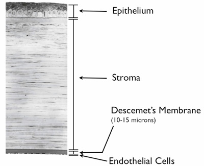
The cornea is the clear front wall of the eye, similar to a watch crystal. Functionally, the cornea has 3 main layers. All layers are clear and must be so to focus light properly.

The outer 10% of the cornea, the epithelium, is the protective skin layer that has sensation. It tells you to blink when your eyes are dry or if you get something in it. The epithelium protects the stroma from infection, scarring, drying out, and other potential harm. Just like the outer layer of your skin, the corneal epithelium sheds and regenerates itself every week. The new cells are grown by corneal epithelial stem cells. Therefore, an eye with an area of bad or opaque epithelium that blocks vision likely does not need a transplant to remove to opacity. Instead of the opacity typically only needs to be removed so that new healthy and clear epithelium can grow in (superficial keratectomy or SK). All other corneal layers do not regenerate and would need to be replaced or transplanted if removed.

The middle layer, the stroma, is the tough structural portion that makes up about 85% of the cornea thickness. The stroma is covered on the outside by the epithelium and on the inside by Descemet’s membrane.

The inner layer of the cornea, Descemet’s membrane, makes up 3 to 5% of its thickness. It is a thin Saran wrap-like membrane that has a single layer of extremely important endothelial cells living on its inner surface. The cornea needs oxygen and nutrients delivered to it, but it does not have blood flow like other parts of the body since blood vessels would make it opaque. The cornea is nourished by a clear fluid called aqueous humor. Aqueous inside the eye is made by arteries and is drained by veins. Descemet’s membrane limits the rate that aqueous humor can flow into the cornea. The endothelial cells pump out “used” aqueous humor so that it does not build up in the cornea which would otherwise make it opaque.

Corneal Layer Diagram

Is it possible to remove and/or transplant just one layer of the cornea?

Yes. We can explain the different types of cornea transplantation using the analogy of a wall. Think of the stroma as the drywall and bricks, Descemet’s membrane and endothelium as the wallpaper, and the epithelium as the temporary protective layers of clear coat that the owner must paint on the bricks from time to time.

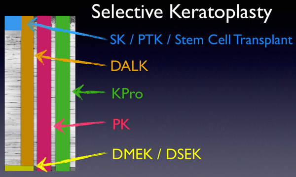
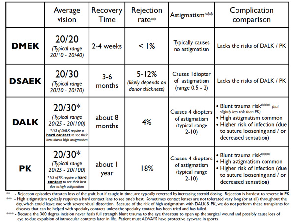
Not that long ago, the only available surgery was a full thickness cornea transplant or penetrating keratoplasty (PK). With PK, all of the corneal layers are transplanted, and new epithelial cells grow over the new cornea in time. The entire wall is knocked out with a hammer, and a new wall and all its layers are brought in. The owner provides a new layer of clear coat over time. A prosthetic corneal transplant (keratoprosthesis (KPro) is an artificial full-thickness corneal transplant.

Selective keratoplasty surgeries are a huge advance over PK in that they allow us to replace just the portion of the cornea that is diseased.

Selective Keratoplasty Diagram

    • Superficial keratectomy (SK) and phototherapeutic keratectomy (PTK) remove the epithelium (and possibly some superficial stroma with PTK) so that new healthier epithelium can grow in. The corneal epithelium is the only layer of the cornea that regenerates. So, SK and PTK aren’t true transplants. They are like removing some imperfections in the layer of clear coat on the outside of the house so that the owner can lay down new clear coat. Conversely, an epithelial stem cell transplant is necessary when the owner’s can of clear coat runs out or goes bad. It becomes necessary to give the owner a new supply of clear coat, which involves transplanting in a special area of a donor cornea.
    • Deep anterior lamellar keratoplasty (DALK) transplants all stroma but leaves the host’s healthy Descemet’s membrane and endothelium behind to allow less risk of rejection or less risk from blunt trauma. The entire wall is changed out, but the host’s own delicate wallpaper is left behind.
  • Descemet’s membrane endothelial keratoplasty (DMEK) and Descemet’s stripping automated endothelial keratoplasty (DSAEK) replace Descemet’s membrane and endothelium without removing the host’s stroma. DMEK is more selective than DSAEK. Both DMEK and DSAEK remove old Descemet’s membrane and endothelium. DMEK adds a new Descemet’s membrane and endothelium only. DSAEK also adds a new Descemet’s membrane and endothelium but with an additional layer of donor stroma. Using the wallpaper analogy, in both DMEK and DSAEK the old wallpaper is removed. With DMEK, only new wallpaper is inserted. With DSAEK, a new piece of drywall that has new wallpaper on it is inserted on top of the old drywall.

Why do we need different types of transplants? Why not just do PK for everyone

PK has many limitations regarding vision, astigmatism, recovery time, rejection rate, and other risks. All types of selective transplants improve the outcomes when compared to PK. DMEK especially has many other benefits compared to DSAEK and PK. For example, performing PK for purely endothelial disease is outdated. It would be like knocking down the wall and bringing in a whole new wall when the only thing needed is new wallpaper!

Cornea Transplant Procedures Diagram

Do I need to go on systemic immunosuppressive medications for a corneal transplant?

Not for most types of corneal transplants. Epithelial stem cell transplants are an exception. When someone gets a kidney or a heart transplant, they need to go on strong medications that make the immune system less aggressive and less likely to attack the transplant. These medications have some potentially significant side effects.

Because there are no blood vessels in the cornea, it is generally invisible to the immune system. Thus for corneal transplants, with the exception of epithelial stem cell transplants, we only need anti-inflammatory eye drops to prevent an immune system attack or rejection. Most patients get down to one drop a day after several few months.

Rarely, patients with aggressive immune systems require stronger systemic medications to prevent rejection. Even with aggressive immune systems, rejection is still very rare with DMEK and is more commonly seen with DSAEK, DALK, or PK. PK has the highest risk of rejection.

After a corneal transplant, do I have to take any precautions if I ever receive a vaccination?

There is some data to suggest that the increase in the immune system activity after vaccination, including the flu and shingles vaccines, may put the graft at a slightly increased risk for a rejection episode. To combat this, we typically recommend that if you were down to just one steroid drop a day, immediately following a vaccine you should increase the steroid drops to four times a day for one week, then two times a day for one week, and then go back to just one daily. If you have previously been told that you cannot take steroid drops due to pressure problems or other issues, ask your cornea surgeon for advice before getting a vaccine. transplant.

How long will my transplant last?

Traditional full thickness corneal transplants (PK) last about 20 years. Cell count studies show that, with the passage of time, transplants still lose endothelial cells gradually just like any other cornea, but usually at a faster rate. When the endothelial cell counts fall low enough, the transplant becomes opaque and fails. Since DMEK and DSAEK are relatively new, it is not possible to say how long they will last; however, preliminary data is encouraging, especially for DMEK. There is variation between transplants, but early data suggest some transplants can even last one’s lifetime. Either way, the replacement of a transplant is possible.

Dr. Tenkman is studying variables that may reveal which donors have cells that are more resistant to death and also surgical techniques that are minimally harmful to endothelial cells. Many surgeons suggest it is normal to lose 30 to 50% of the donor’s endothelial cells during surgery. We have some early data suggesting less than 10% cell loss from surgery when selecting a specific subset of donors.

Does a rejection episode mean that I will lose my cornea transplant?

No. If untreated, a rejection episode can cause significant damage. But usually, the episode stops with an increase in anti-inflammatory eye drops. Patients are instructed to RSVP: come see us if they develop Redness, Sensitivity to light, Vision reduction, or Pain. It’s key to see us right away for prompt diagnosis and treatment.

Will I need glasses after my transplant? Could laser vision correction be done to reduce any postoperative need for glasses?

Whether or not you need glasses after your transplant depends on many factors. DMEK transplants reduce glasses dependence the most often. DSAEK is less predictable. DALK and PK can both frequently cause high astigmatism that needs glasses or even hard contacts to attain good vision.

Some patients are candidates for ASA (advanced surface ablation) to minimize their dependence on glasses or contacts after corneal transplant surgery. ASA is a laser vision correction procedure similar to LASIK. Whether or not ASA could be done to reduce dependence on glasses or contacts after surgery depends on several factors and is taken on a case by case basis.

What are the costs and risks of a cornea transplant?

Corneal transplant tissue is donated, but still typically costs $3,700 or more. The fees go to the eye banks that must harvest the corneas, screen for possible diseases that could be transferred to the patient, transport, and process the corneas, and deliver them to the surgery center… all within just a few days. The cost of receiving a transplant includes not just the tissue, but the surgery center, surgeon, and anesthesia fees. Fortunately, these costs are less at an outpatient surgery center and are typically covered by Medicare and private insurance, although patients may be responsible for deductibles and co-payments. Generally, DMEK and DSAEK can be performed in an outpatient surgery center whereas DALK and PK more often have to be done at a hospital. Having surgery at a hospital can increase costs several-fold.

The risk of infection from a corneal transplantation procedure is about the same as following a routine blood transfusion. The risk of receiving a disease from a transplant is very low.

Other risks include primary graft failure, rejection, graft dislocation, cataract formation, glaucomainfection, irregular astigmatism, double vision, bleeding, iris damage, vitreous prolapse, and cystoid macular edema. Most of these complications are quite rare but are theoretically possible with all types of eye surgery. Patients using steroid drops, which all corneal transplants require, have to be followed for the development of increased pressure. High eye pressure usually gives no symptoms, so follow up is crucial to allow the doctor to treat as necessary with medication or procedures. Patients are usually seen every 3 to 6 months for life depending on the strength of steroid used.

In our hands, the overall success rate for a DMEK or DSAEK graft attaching and working well is about 99%. Should the transplant fail due to rejection or otherwise, it would need to be repeated.

Because the time to heal is longer for DALK and PK, and because they involve more risk, the odds of a DALK or PK failing to restore vision is more significant than for DMEK or DSAEK. DALK and PK have several additional risks.

  • DALK, and more so PK, have a higher risk of bleeding during surgery while the patient’s own cornea is removed and the eye is depressurized. Although not common, such bleeding can do significant damage to the eye.
  • DALK, and more so PK, also have a significant risk of rejection. Higher doses of steroid drops may be necessary in here to prevent or treat rejection, which can increase the risk of increased eye pressure (glaucoma).
  • DALK and PK involve a 360-degree corneal incision. Such large corneal wounds don’t heal with full strength and are at risk to split open with moderate blunt trauma (ie if a patient fell and hit their face in the shower). If the wound were to split open, it could lead to loss of the eye.
  • DALK and PK require about 16 sutures. These sutures are not removed for many months or even over a year. The sutures are buried in a manner so they cause no pain to the patient. However, they commonly become loose and can cause a foreign body or sandy sensation. More significantly, a loose suture is a risk for infection (corneal ulcer). Corneal ulcers can be more serious in the setting of a corneal transplant because the immune system in the area is suppressed by steroid eye drops.
  • DALK and PK commonly heal with large amounts of astigmatism. The normally spherical corneal surface becomes like an irregularly shaped egg. Since the smooth corneal shape largely determines focus, an irregular cornea causes blurred vision. Corneal astigmatism is measured in diopters. In routine patients who have not had had a corneal transplant, we consider 1 diopter mild astigmatism, 2 diopters moderate astigmatism, and 3 diopters high astigmatism. DALK and PK average about 4 to 5 diopters of astigmatism. A common range is 2 to 10 diopters of astigmatism. Regarding astigmatism risk, there are controllable and uncontrollable causes. The surgeon can try to sew the graft as evenly as possible to reduce risk of severe astigmatism; however, the surgeon cannot control the natural tension lines in the cornea or how symmetrically the tension lines will balance as the wound heals. Severe or irregular astigmatism cannot be corrected with glasses. Special hard contact lenses are often necessary. On average, one-third of all DALK and PK patients need hard contact to see their best due to astigmatism. Therefore, at Bennett & Bloom, we never perform DALK or PK for keratoconus patients to try to get out of contact lenses. The patient could go through all the added cost and risk of surgery only to end up where they started… or worse. DALK and PK are reserved for severe disease that cannot be fixed by contact lens wear


For corneal transplant at Bennet & Bloom Eye center in the USA or for medical tourism in the USA, contact Eyeupdate Clinic & Optical Supplies, 01 Ajuwon junction, Off Elliot bus stop, Iju Ishagah, Lagos.Tel: +2347030000001, +19093663551

Muscles, Nerves, and Blood Vessels in the Human Eye

By David Terfera, Shereen Jegtvig

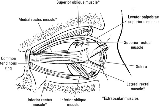
Muscles enable you to move your eyes. Ocular nerves allow you to interpret what you see and blood vessels keep your eyes oxygenated. Six muscles, collectively called the extraocular muscles, move the eyeball. A seventh muscle moves the eyelid and is also found in the orbit.

The muscles of the human eye

The following muscles help your eyes move around.

image0.jpg

  • Levator palpebrae superioris: Originates on the sphenoid bone above the optic canal. It inserts into the superior tarsis and skin of the eyelid. It’s innervated by the oculomotor nerve and elevates the superior eyelid.

  • Superior oblique: Originates on the sphenoid bone and inserts into the sclera deep to the superior rectus muscle. It’s innervated by the trochlear nerve and abducts, depresses, and medially rotates the eyeball.

  • Inferior oblique: Originates on the anterior part of the orbital floor and inserts onto the sclera deep to the lateral rectus muscle. It’s innervated by the oculomotor nerve and abducts, elevates, and laterally rotates the eyeball.

  • Superior rectus: Originates on the common tendinous ring and inserts into the sclera behind the corneoscleral junction. It’s innervated by the oculomotor nerve, and it elevates, adducts, and medially rotates the eyeball.

  • Inferior rectus: Originates on the common tendinous ring and inserts into the sclera behind the corneoscleral junction. It’s innervated by the oculomotor nerve and depresses, adducts, and laterally rotates the eyeball.

  • Medial rectus: Originates on the common tendinous ring and inserts into the sclera behind the corneoscleral junction, this muscle is innervated by the oculomotor nerve and adducts the eyeball.

  • Lateral rectus: Originates on the common tendinous ring and inserts into the sclera behind the corneoscleral junction. It’s innervated by the abducent nerve and abducts the eyeball.

The nerves of the eye

The eyes are served by the following cranial nerves and their branches:

image1.jpg

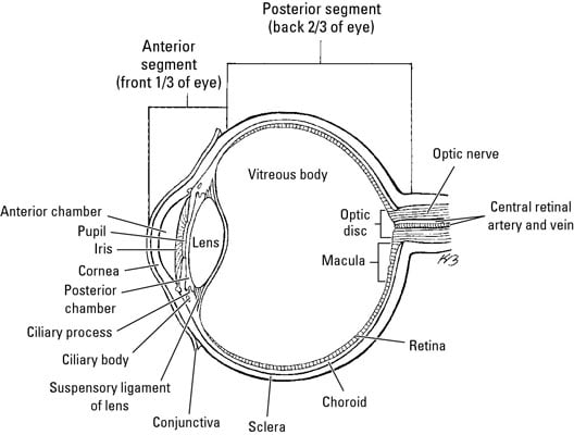
  • Optic nerve (CN II): Sensory nerve that transmits impulses from the retina to the brain

    • Oculomotor nerve (CN III), trochlear nerve (CN IV), and abducent nerve (CN VI): Enter the orbital space through the superior orbital fissure to innervate the extraocular muscles.

    • Ophthalmic nerve (part of the trigeminal nerve, CN V): This nerve has three branches:

      • The lacrimal nerve runs to the lacrimal gland and gives off branches to the conjunctiva and skin of the superior eyelid.

      • The frontal nerve enters through the superior orbital fissure and provides sensory innervation to the superior eyelid, scalp, and forehead.

      • The nasociliary nerve is the sensory nerve to the eyeball. It also has branches that serve the orbit and other parts of the face. One of its branches, the infratrochlear nerve, supplies the eyelids, conjunctiva, and lacrimal sac.

    • Ciliary ganglion: This group of postsynaptic parasympathetic nerve cell bodies is associated with the oculomotor nerve and ophthalmic nerve (CN V1). Presynaptic parasympathetic fibers from the oculomotor nerve synapse on the cell bodies of postsynaptic parasympathetic neurons in the ciliary ganglion.

      Short ciliary nerves emerge from the ciliary ganglion and enter the eye. The short ciliary nerves contain postsynaptic parasympathetic fibers from the ciliary ganglion, afferent fibers of the nasociliary nerve, and postsynaptic sympathetic fibers from the internal carotid plexus. Postsynaptic parasympathetic fibers innervate the ciliary muscle and sphincter pupillae muscle. Afferent fibers convey sensory impulses from the iris and cornea. Postsynaptic sympathetic fibers innervate the dilator pupillae muscle.

      The long ciliary nerves contain afferent and postsynaptic sympathetic fibers from the nasociliary nerve. Long ciliary nerves bypass the ciliary ganglion and run to the iris, cornea, and dilator pupillae muscle.

    The blood vessels

    Blood flow to the orbit (and beyond) comes from branches of the internal carotid artery, chiefly via the ophthalmic artery and its branches:

    • Ophthalmic artery: Branches from the internal carotid artery and passes through the optic canal into the orbital cavity

    • Central artery of the retina: Runs from the ophthalmic artery to the eyeball alongside the optic nerve; it branches at the optic disc and supplies the retina

    • Supraorbital artery: Starts at the ophthalmic artery and exits the orbit at the supraorbital notch to supply the forehead and scalp

    • Supratrochlear artery: Runs from the ophthalmic artery to the forehead and scalp

    • Lacrimal artery: Runs from the ophthalmic artery along the lateral rectus muscle to supply the lacrimal gland, conjunctiva, and the eyelids

    • Dorsal nasal artery: Branches from the ophthalmic artery and runs along the nose to supply it with blood

    • Short posterior ciliary arteries: Branch from the ophthalmic artery and pierce the sclera at the edge of the optic nerve; they supply the choroid and the rods and cones of the retina

    • Long posterior ciliary arteries: Branch from the ophthalmic artery and pierce the sclera to supply the ciliary body and iris

    • Posterior ethmoidal artery: Leaves the ophthalmic artery to supply blood to ethmoidal cells

    • Anterior ethmoidal artery: Runs from the ophthalmic artery to supply ethmoidal cells, frontal sinus, nasal cavity, and skin over the nose

    • Anterior ciliary artery: Runs from the muscular branches of the ophthalmic artery through the sclera near the rectus muscles and forms an arterial network in the iris and ciliary body

    • Infraorbital artery: Runs from the maxillary artery along the infraorbital groove and out to the face

    Blood is returned from the orbits via the superior and inferior ophthalmic veins, which pass through the superior orbital fissure into the cavernous sinus. The central vein of the retina may join an ophthalmic vein or enter the cavernous sinus directly. Vorticose veins drain the vascular layer of the eyeball, and the scleral venous sinus encircles the anterior chamber of the eyeball.

How alcohol affects your eyes -Eye care perspective

Although light consumption of alcohol probably won’t cause any health problems, drinking alcohol excessively can have harmful effects on your body, including your eyes. Heavy drinking of alcohol may cause problems with your vision and overall eye health including the following:

    • Decreased visual performance: Your overall visual performance may be altered since drinking heavily impairs brain function. You may have blurred vision or double vision due to weakened eye muscle coordination. You may also experience delayed reactions while driving.
    • Slow pupil reactions: Alcohol tends to affect the speed at which your iris constricts and dilates. A driver that has been drinking alcohol cannot adapt as quickly to oncoming headlights.
    • Decreased peripheral vision: Drinking alcohol has also been shown to decrease the sensitivity of your peripheral vision. This may give you the effect or perception of having tunnel vision.
  • Decreased contrast sensitivity: Drinking too much alcohol can alter your contrast sensitivity, or how precise you can discern between shades of gray. Driving in rain or fog will be much more dangerous.
  • Optic neuropathy: Also referred to as tobacco-alcohol amblyopia, people who drink or smoke in excess can develop optic neuropathy. You might develop a painless loss of vision, decreased peripheral vision or reduced color vision. Even though studies have shown the vision loss to be a result of a nutritional deficiency, some professionals believe that the condition develops because of toxic effects of alcohol and tobacco.
  • Frequent migraines: Alcohol has been shown to be a trigger for severe migraine headaches in some people. You may experience a temporary, but debilitating visual aura before the onset of the headache. The visual aura may appear as blind spots, graying of vision or zig-zag patterns of light.​
  • Poor cosmetic appearance: Drinking can cause eye redness. Alcohol causes the blood vessels in your eyes to expand, making them more prominent.


If you have any issues with your eyes, visit or call : Eye update Eye clinic & optical supplies, 01, Ajuwon junction, Ajuwon bus stop, Akute/Ajuwon road, off Elliot bus stop, Iju-Ishagah. Tel: 08034971582

Postal Codes of Streets in Ojodu Lagos Nigeria

Ojodu Streets Zip Codes

Address:
Area: Ojodu, Lagos, Lagos State. Associated Zip Code: 100213

District Name: Area: Ojodu
Local Government Area: Lagos
State: Lagos State
Associated Zip Code: 100213

Streets: (260)
Street name Zip Code
Raimi St St. 100213
Rasheed Baruwa St. 100213
Regina Ola Ore St St. 100213
Sogo St St. 100213
Sowebo Oladipupo St. 100213
Soyemi St St. 100213
Sule Abore St St. 100213
Taiwo Ishola St. 100213
Taiwo Okolu St. 100213
Tijani Bello St St. 100213
Unity Cl St. 100213
Zinzon S St. 100213
Alara St St. 100213
Adesina St St. 100213
Funsho St St. 100213
Tobun St St. 100213
Olumo St St. 100213
OlasimboSt St. 100213
Olu Osifeso St St. 100213
Remi Abuah St St. 100213
Majaro St St. 100213
Akintunde St St. 100213
Mobolaji St St. 100213
Johnson St St. 100213
Araromi St St. 100213
Ajayi St St. 100213
Aderibigbe St St. 100213
Lawal St St. 100213
Adebiyi St St. 100213
Abiodun St St. 100213
Adesina St St. 100213
Barikisu Iyade St St. 100213
University Rd St. 100213
Davies St St. 100213
Ayodele St St. 100213
Amusa St St. 100213
Sule St St. 100213
Abudu St St. 100213
Amen St St. 100213
Ajayi Bembe St St. 100213
Obadina St St. 100213
Mosuro St St. 100213
Sadiku St St. 100213
Ebun St St. 100213
Balogun St St. 100213
Are Ago St St. 100213
Abule Ijesha Rd St. 100213
Morris Rd St. 100213
Eletu Odibo St St. 100213
Shodipe St St. 100213
Saka Ln St. 100213
Martins St St. 100213
Odenike St St. 100213
Shobowale St St. 100213
Otun St St. 100213
Oduntan St St. 100213
Oguntuga St St. 100213
Silver St St. 100213
Bailey St St. 100213
Akinsola St St. 100213
Bamigbopa St St. 100213
Alaka St St. 100213
Mosuro St St. 100213
Bankole St St. 100213
Lemboye St St. 100213
Ogabi St St. 100213
Iwaya Rd St. 100213
Owodunni St St. 100213
Memuda St St. 100213
Ogunkoya St St. 100213
Abiye St St. 100213
Ijebu Quarters St. 100213
Oloto St St. 100213
Victoria St St. 100213
Ajoke St St. 100213
Aderupoko St St. 100213
Imoru St St. 100213
Audu Bale St St. 100213
Adegbenro St St. 100213
Salami St St. 100213
Church St St. 100213
Akanni St St. 100213
Omitogun St St. 100213
Yey St St. 100213
Ogunbiyi St St. 100213
Omotola St St. 100213
Arowolo St St. 100213
Balogun St St. 100213
Moshalashi St St. 100213
Akintola St St. 100213
Owode St St. 100213
Pedro St St. 100213
Omiaro St St. 100213
Ogbwe St St. 100213
Oyewale St St. 100213
Onituere St St. 100213
Ogundimu St St. 100213
Joseph Marrison St St. 100213
Gbede St St. 100213
Dacosta St St. 100213
Ogayemi St St. 100213
Afolabi Ayorinde St St. 100213
Wugbo St St. 100213
Yovi St St. 100213
Akinlehinb St St. 100213
Ayato St St. 100213
Oke Anaiye St St. 100213
Igbore St St. 100213
Development St St. 100213
ALh. Rainmi St. 100213
Araromi St St. 100213
Iman Ln St. 100213
Wright St St. 100213
Makoko Rd St. 100213
Dany Est St. 100213
Akinbo St St. 100213
Erejuwa St St. 100213
Ramotu St St. 100213
Olaiya St St. 100213
Church St St. 100213
Ajelekoko St St. 100213
Igbehin Adun St St. 100213
Abudu St St. 100213
Rafiu St St. 100213
Adetia St St. 100213
Olulu St St. 100213
Olumide St St. 100213
Sariyu St St. 100213
Obalomu St St. 100213
Falodun St St. 100213
Olayide St St. 100213
Alafia St St. 100213
Oluwatoyin St St. 100213
Bale St St. 100213
Alh.Sule St St. 100213
Alh. Raimi St. 100213
St Finbar College St St. 100213
Moronfolu St St. 100213
Community Rd St. 100213
Obayan St St. 100213
Amos Bakare St St. 100213
Afolabi Brown St St. 100213
Funmilayo Oyaronke St St. 100213
Tunde Bello St St. 100213
Emily Akinola St St. 100213
Iyiola Olawale St St. 100213
AdeyinkaOsijo St St. 100213
Abdulahi St St. 100213
Wulemotu Ajoke St St. 100213
Abayomi St St. 100213
Olarewaju St St. 100213
Rikett Cl St. 100213
Ayetoro St St. 100213
Oyenuga St St. 100213
Oridami St St. 100213
Itesiwaju St St. 100213
Sholanke St St. 100213
Shuwu Court St. 100213
Mafowoku St St. 100213
Adenuga Kajaro St St. 100213
Ojo St St. 100213
Abiodun Cl St. 100213
Bandi Adewale St St. 100213
Olorunjare St St. 100213
Ayinde St St. 100213
Onafeko St St. 100213
Adetayo Osho St St. 100213
Osanyinpeju St St. 100213
Abatan Rd Rd. 100213
Alonge St St. 100213
Abigi St St. 100213
Awolaja St St. 100213
Abiodun St St. 100213
Oluwole St St. 100213
Adebisi Awosoga 100213
Animashaun St St. 100213
Adebolawale St St. 100213
Adegoke Ajayi St St. 100213
Ademola Ajasa 100213
Adeniyi Oke St St. 100213
Adewale 100213
Adewale Adegun 100213
Adeyemo Akapo 100213
Adisa Ambali Cl Cl. 100213
Agoro 100213
Aina St St. 100213
Ajana Cl Cl. 100213
Ajayi St St. 100213
Akin Oladeguro 100213
Akinola St St. 100213
Akinsanya St. 100213
Akiode Cr St. 100213
Alh. Abass St St. 100213
Alh. Ashafa St St. 100213
Alh.Bakare St. 100213
Alh. Bashiru St St. 100213
Ali Baogun St. 100213
Animashaun St St. 100213
Anu Oluwapo St St. 100213
Ayo Alabi St St. 100213
Babatunde Ladega St. 100213
Bakare St St. 100213
Bamako St St. 100213
Bank Shonibare St St. 100213
Banni St St. 100213
Basiaru St St. 100213
Beatrice Spencer St. 100213
Bisi Gbadina St St. 100213
Bosude St St. 100213
Bunuyo Cl St. 100213
Burdland St St. 100213
College Rd St. 100213
Arogangan St. 100213
Debo Aina St. 100213
Efon Alaye St. 100213
Foly St St. 100213
Gbenga Kilo St St. 100213
Gbite St St. 100213
Gbolahon Owolabi Cl St. 100213
Ibadan Ewy St. 100213
Ibrahim Adekunle St St. 100213
Ifo Cl St. 100213
Irepodun St St. 100213
Isale Tapa St St. 100213
Isheri Holiday Inn St St. 100213
Isheri Rd St. 100213
Ishola Bello St St. 100213
Ivern Ave St. 100213
Jacob Runsance Cl St. 100213
Jide Ayo St. 100213
Kadiri St. 100213
Kayode Ali Cl St. 100213
Kupoluyi St St. 100213
LSDPC Quarters St. 100213
Lola Holloway St. 100213
Mafaosky Cl St. 100213
Moses Adebayo St St. 100213
Muyibo St St. 100213
Muyiwa Oyefusi St. 100213
New Isheri Rd St. 100213
Oba Bobington Ashoye St. 100213
Obafolabi St St. 100213
Oboku St St. 100213
Odozi St St. 100213
Ogunbiyi Akande St. 100213
Ola Oluwa St St. 100213
Oladipupo Oluwole St St. 100213
Olajide Ave St. 100213
Olaleke Taiwo St St. 100213
Olanipekun St. 100213
Olatunde Badmus St. 100213
Olawale St. 100213
Olumuyiwa St. 100213
Oluwadamilola Falade St. 100213
Oluwatoyin Cl St. 100213
Omofade Cr St. 100213
Omorogie St St. 100213
Orimeta St St. 100213
Popoola Oyinloye Cl St. 100213
Popola St St. 100213

Chalazion -Eye/Optical clinic near Ojokoro

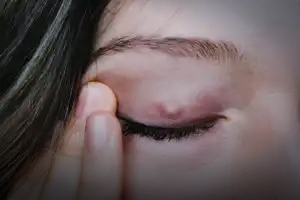
chalazion is a small, usually painless, lump or swelling that appears on your eyelid. A blocked meibomian or oil gland causes this condition. It can develop on the upper or lower eyelid, and may disappear without treatment. Chalazia is the term for multiple chalazion.

A chalazion is sometimes confused with an internal or external stye. An internal stye is an infection of a meibomian gland. An external stye is an infection in the area of the eyelash follicle and sweat gland. Styes are usually painful and chalazia usually aren’t. Chalazia may develop after styes.

You should see your eye doctor if you think you have a chalazion, especially if it blocks your vision or if you’ve had chalazia in the past.

Causes and risk factors

The chalazion is caused by a blockage in one of the tiny meibomian glands of the upper and lower eyelids. The oil these glands produce helps to moisten the eyes.

Inflammation or viruses affecting the meibomian glands are the underlying causes of chalazia.

Chalazia are more common in people with inflammatory conditions like seborrheaacnerosacea, chronic blepharitis, or long-term inflammation of the eyelid. They’re also more common in people with viral conjunctivitis or an infection covering the inside of the eyes and eyelids.

Recurring or unusual chalazia may be symptoms of more serious conditions, but these are rare.

Symptoms

A chalazion usually appears as a painless lump or swelling on your upper or lower eyelid. Chalazia may affect both upper and lower lids and can occur in both eyes at the same time. Depending on the size and location of the chalazion, it may blur or block vision.

Although not as common, a chalazion may be red, swollen, and painful if an infection is present.

Diagnosis

In most cases, a doctor can diagnose this condition by taking a close look at the lump on your eyelid. Your doctor will also ask about your symptoms to determine if the lump is a chalazion, a stye, or something else.

Treatment

Some chalazia can go away without treatment. If your doctor does recommend treatment, options may include:

Home care

First, do not try to squeeze the chalazion. It’s best if you touch it as little as possible.

Instead, you should apply a warm compress to your eyelid four times per day for about 10 minutes at a time. This can reduce the swelling by softening the oils in the blocked gland. Make sure you wash your hands before you touch the area.

Your doctor may also tell you to gently massage the lump a few times per day or to scrub your eyelid. Your doctor may also prescribe eye drops or eyelid creams.

Medical treatment

If the chalazion doesn’t go away with home treatment, your doctor may recommend a corticosteroid injection or a surgical procedure. Both the injection and the surgery are effective treatments.

The choice of treatment depends on several different factors. Your doctor will explain the benefits and risks.

Preventing a chalazion

It’s not always possible to avoid getting a chalazion. This is especially true if you’re prone to this type of eye problem. But there are a few things that you can do to prevent this condition:

  • Always wash your hands before touching your eyes.
  • Make sure that anything that comes in contact with your eyes, such as contact lenses and glasses, is clean.
  • If you have a condition that increases your chance of developing chalazia, follow your doctor’s instructions to help control them.


If you have any eye infection or discomfort with the eyes, call or visit:

Eye update Eye clinic & opticals 01, Ajuwon junction, Ajuwon bus stop, beside BPNL filling station, Akute/Ajuwon road, off Elliot bus stop, Iju-Ishagah Near Ojokoro. Tel: 0803497158, 08107531046