Uveitis

Overview

Uveitis is a form of eye inflammation. It affects the middle layer of tissue in the eye wall (uvea).

Uveitis (u-vee-I-tis) warning signs often come on suddenly and get worse quickly. They include eye redness, pain and blurred vision. The condition can affect one or both eyes, and it can affect people of all ages, even children.

Possible causes of uveitis are infection, injury, or an autoimmune or inflammatory disease. Many times a cause can’t be identified.

Uveitis can be serious, leading to permanent vision loss. Early diagnosis and treatment are important to prevent complications and preserve your vision.

Symptoms
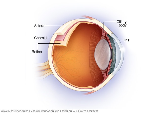
Uvea where uveitis occursEye with uveaOpen pop-up dialog box

The signs, symptoms and characteristics of uveitis may include:

  • Eye redness
  • Eye pain
  • Light sensitivity
  • Blurred vision
  • Dark, floating spots in your field of vision (floaters)
  • Decreased vision

Symptoms may occur suddenly and get worse quickly, though in some cases, they develop gradually. They may affect one or both eyes. Occasionally, there are no symptoms, and signs of uveitis are observed on a routine eye exam.

The uvea is the middle layer of tissue in the wall of the eye. It consists of the iris, the ciliary body and the choroid. When you look at your eye in the mirror, you will see the white part of the eye (sclera) and the colored part of the eye (iris).

The iris is located inside the front of the eye. The ciliary body is a structure behind the iris. The choroid is a layer of blood vessels between the retina and the sclera. The retina lines the inside of the back of the eye, like wallpaper. The inside of the back of the eye is filled with a gel-like liquid called vitreous.

The type of uveitis you have depends on which part or parts of the eye are inflamed:

  • Anterior uveitis affects the inside of the front of your eye (between the cornea and the iris) and the ciliary body. It is also called iritis and is the most common type of uveitis.
  • Intermediate uveitis affects the retina and blood vessels just behind the lens (pars plana) as well as the gel in the center of the eye (vitreous).
  • Posterior uveitis affects a layer on the inside of the back of your eye, either the retina or the choroid.
  • Panuveitis occurs when all layers of the uvea are inflamed, from the front to the back of your eye.

When to seek medical advice

Contact your doctor if you think you have the warning signs of uveitis. He or she may refer you to an eye specialist (ophthalmologist). If you’re having significant eye pain and unexpected vision problems, seek immediate medical attention.

Causes

In about half of all cases, the specific cause of uveitis isn’t clear, and the disorder may be considered an autoimmune disease that only affects the eye or eyes. If a cause can be determined, it may be one of the following:

  • An autoimmune or inflammatory disorder that affects other parts of the body, such as sarcoidosis, ankylosing spondylitis, systemic lupus erythematosus or Crohn’s disease
  • An infection, such as cat-scratch disease, herpes zoster, syphilis, toxoplasmosis or tuberculosis
  • Medication side effect
  • Eye injury or surgery
  • Very rarely, a cancer that affects the eye, such as lymphoma

Risk factors

People with changes in certain genes may be more likely to develop uveitis. Cigarette smoking has been associated with more difficult to control uveitis.

Complications

Left untreated, uveitis can cause complications, including:

  • Retinal swelling (macular edema)
  • Retina scarring
  • Glaucoma
  • Cataracts
  • Optic nerve damage
  • Retinal detachment
  • Permanent vision loss

Prosthetic Contact Lenses

Prosthetic contact lenses are prescribed to mask flaws and improve the appearance of an eye disfigured from a birth defect, trauma, or eye disease.

If certain structures of the injured or disfigured eye also fail to function properly, special prosthetic lenses can be designed to block excess light from reaching the back of the eye to reduce glare and increase comfort.

Your eye care practitioner can match prosthetic contact lenses to the appearance of a healthy eye by using a pre-made trial set or by ordering custom-painted contact lenses.

Like regular contact lenses, prosthetic contacts can be made of gas permeable or soft lens materials. And most prosthetic lenses can be cleaned and disinfected using the same multipurpose contact lens solutions recommended for conventional soft or gas permeable lenses.

Custom prosthetic lenses can be made in a wide variety of lens powers to correct nearsightednessfarsightedness and/or astigmatism, or they can be produced without corrective power to simply provide a more natural appearance to a blind or disfigured eye.

Eye Conditions That May Benefit From Prosthetic Contact Lenses

Many injuries, conditions and infections can lead to disfigurement of an eye and make an individual feel self-conscious about his or her appearance. Prosthetic lenses can be specially designed to match the appearance of the unaffected eye as closely as possible and make the disfigured eye less conspicuous.

Disfiguring conditions frequently treated with prosthetic contact lenses include incomplete formation of the pupil (aniridia), lack of pigment or color in the eye (albinism) and damage to the cornea from trauma.

People with albinism or other eye conditions creating light sensitivity (photophobia) can benefit from prosthetic lenses that reduce the amount of light entering the eye. Prosthetic lenses also are sometimes used to eliminate double vision (diplopia) caused by certain eye conditions.

Special prosthetic colored contact lenses sometimes are used to improve vision in cases of childhood amblyopia. Two identical-appearing colored contacts are worn, but one lens has an opaque pupil painted on it to block (occlude) light from entering the eye. This lens is worn on the eye with normal sight, forcing greater use of the amblyopic eye to improve vision development in the affected eye.

Occlusion therapy with prosthetic contact lenses can be more effective for treating amblyopia than the use of an eye patch, which children often resist or remove due to embarrassment. But amblyopia treatment with contacts is more expensive due to the cost of the prosthetic lenses.

How Prosthetic Contact Lenses Are Prescribed

The first step in being fit with prosthetic contact lenses is to have a comprehensive eye exam. During this exam, your optometrist or ophthalmologist will thoroughly examine your eyes to rule out conditions that might interfere with successful prosthetic contact lens wear.

The second step is to undergo a contact lens fitting. In addition to performing the steps in a normal contact lens exam and fitting, your eye doctor may also take close-up photographs of your eyes. These color photos are sent to the prosthetic contact lens manufacturer to help the company create a prosthetic lens that matches your natural eye color and appearance as closely as possible.

Some eye doctors who specialize in contacts for hard-to-fit eyes also may have fitting sets of sample prosthetic lenses on hand so you can get an immediate idea of how effectively the lenses can restore a natural appearance to your affected eye(s).