Two Patients With Macular Degeneration Get Artificial Retinas

LYON, France — A 72-year-old woman with dry age-related macular degeneration (AMD) was the first patient to receive an artificial retina implant as part of the multicenter PRIMAvera clinical trial, which is looking at the safety and efficacy of the PRIMA system.

“Her vision was severely impaired by this condition. We used the ETDRS chart to assess her visual acuity, as this is the current method in ophthalmology. She was able to read only nine letters,” said Laurent Kodjikian, MD, PhD, from Hôpital de la Croix-Rousse in Lyon, who is a former president of the French Society of Ophthalmology. To put that in perspective, a person with normal vision can make out 85 letters, he explained.

“The goal is to get her reading another ten letters,” he told Medscape Medical News.

The hospital where Kodjikian works is one of six centers in France selected to take part in the ongoing AMD clinical trial; other study sites are in Germany, Italy, the Netherlands, and Spain. The 38 study participants will be followed for 12 months after implantation to assess visual acuity and adverse events, and outcomes will be monitored for 3 years. Investigators hope that the findings will lead to the device receiving authorization to enter the market.

The PRIMA artificial retina system has three elements: a tiny wireless retinal implant; a pair of glasses with a camera and digital projector; and a portable processor connected to the projector. The camera captures visual scenes from the surrounding environment. The processor uses algorithms to process and simplify the images, which are then sent back to the glasses. The digital projector uses pulses of infrared light to project the processed images onto the retinal implant’s photovoltaic receptors. These receptors then convert the optical information into electrical stimulation, which excites the nerve cells of the inner retina, allowing them take in the information and transmit it, via the optic nerve, to the brain. This then induces visual perception.

A Delicate Operation

To implant the chip, Kodjikian made a rather large incision — 3.5 mm — and then peeled off the retina, all while looking through a surgical microscope.

“It’s easy to peel off the retina in a healthy eye. However, the procedure becomes more difficult in an eye affected by dry AMD, where the retina tissue is not only very thin and firmly attached to the back wall of the eye, but is also very fragile. A lot can go wrong during this step, so we have to really take our time,” he said. “You can’t go too deep, and if you go too close to the surface, you risk perforating the retina. Like walking on a very thin tightrope, there’s danger all around, and very little room for error.”

After the chip was inserted under the retina, Kodjikian put the retina tissue back into place. “This was the first time I’d ever done this kind of procedure, and it was quite a challenge,” he told Medscape.

The operation took 2.5 hours, which is much less time than the 4 to 5 hours estimated by the manufacturer.

The patient will undergo rehabilitation for 12 months to help her adapt to the system. “Our hope is that this patient will be able to see better with the implant. She probably won’t get to the point of being able to drive a car. And while reading novels in small print may not be possible, it’s quite likely that she’ll be able to read large-print editions,” Kodjikian explained.

Upon activation of the retinal prosthetic, the patient experienced visual impressions that she couldn’t see before the surgery. And 1 month after the procedure, things seem to be on track, according to a press release. “The postoperative result is excellent. There are no complications, the chip is perfectly in place and the vision has not been degraded by the operation. She should now start to improve thanks to rehabilitation,” said Kodjikian.

“Of the various artificial retina systems out there, this one is the most sophisticated because it has the most pixels. The technology will certainly continue to advance. But for the time being, the clinical study should allow us to show that it does work,” he concluded.

In December 2021, he implanted an artificial retina in a second patient, and it took him 50 minutes less than the first one.

This article originally appeared in the French edition of Medscape.

The authors have disclosed no relevant financial relationships.

Eyeupdate Eye-clinic & Optical Supplies, p1, Ajuwon bus stop, Akute/Ajuwon Road, Ajuwon (beside BPNL/MRS Filling station).
Tel: 08107531046, 08034971582.

Our eye-clinic/optical care services cover people from the following locations and near you:
alagbole, akute, ajuwon, lambe, matogun, matogbun, giwa, okearo, koye, oke, arifanla, baale, Road, fagba, abule, egba, agege, ogba, ikeja, G. R. A, oshodi, Berger, ojodu, omole, magodo, yaba, mowe, ibafo, shagamu, Ishagah, shomolu, heritage, estate, ajao, College Road, iju, Road, water, works, omole, magado, ketu, ojota, ikoyi, akiode bus stop, grammar school, first gate, Ala Hausa, Yoruba, Igbo, Benin, Edo, Association, Club, Ikorodu, Ketu, ijoko, jankara, Ladipo, Ojota, Shomolu, Cele, Ogun, Lagos, Lagos Island, Lagos mainland, Ifako, Ijaye, eti osa, Oyo, Lisa, Obawole,Iju, Ishagah, Ishaga, Isaga, Isagah, Ishasi,Obawale,Owode, sang ota, abule egba, redeem camp, Ijebu ode, toll gate, bus stop, Street, along, Nigeria.

Uveitis

Overview

Uveitis is a form of eye inflammation. It affects the middle layer of tissue in the eye wall (uvea).

Uveitis (u-vee-I-tis) warning signs often come on suddenly and get worse quickly. They include eye redness, pain and blurred vision. The condition can affect one or both eyes, and it can affect people of all ages, even children.

Possible causes of uveitis are infection, injury, or an autoimmune or inflammatory disease. Many times a cause can’t be identified.

Uveitis can be serious, leading to permanent vision loss. Early diagnosis and treatment are important to prevent complications and preserve your vision.

Symptoms

Uvea where uveitis occursEye with uveaOpen pop-up dialog box

The signs, symptoms and characteristics of uveitis may include:

  • Eye redness
  • Eye pain
  • Light sensitivity
  • Blurred vision
  • Dark, floating spots in your field of vision (floaters)
  • Decreased vision

Symptoms may occur suddenly and get worse quickly, though in some cases, they develop gradually. They may affect one or both eyes. Occasionally, there are no symptoms, and signs of uveitis are observed on a routine eye exam.

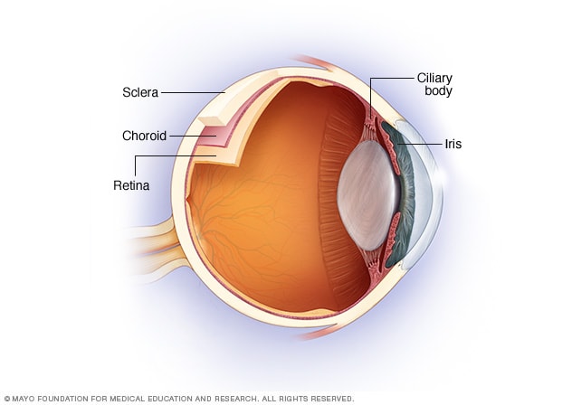
The uvea is the middle layer of tissue in the wall of the eye. It consists of the iris, the ciliary body and the choroid. When you look at your eye in the mirror, you will see the white part of the eye (sclera) and the colored part of the eye (iris).

The iris is located inside the front of the eye. The ciliary body is a structure behind the iris. The choroid is a layer of blood vessels between the retina and the sclera. The retina lines the inside of the back of the eye, like wallpaper. The inside of the back of the eye is filled with a gel-like liquid called vitreous.

The type of uveitis you have depends on which part or parts of the eye are inflamed:

  • Anterior uveitis affects the inside of the front of your eye (between the cornea and the iris) and the ciliary body. It is also called iritis and is the most common type of uveitis.
  • Intermediate uveitis affects the retina and blood vessels just behind the lens (pars plana) as well as the gel in the center of the eye (vitreous).
  • Posterior uveitis affects a layer on the inside of the back of your eye, either the retina or the choroid.
  • Panuveitis occurs when all layers of the uvea are inflamed, from the front to the back of your eye.

When to seek medical advice

Contact your doctor if you think you have the warning signs of uveitis. He or she may refer you to an eye specialist (ophthalmologist). If you’re having significant eye pain and unexpected vision problems, seek immediate medical attention.

Causes

In about half of all cases, the specific cause of uveitis isn’t clear, and the disorder may be considered an autoimmune disease that only affects the eye or eyes. If a cause can be determined, it may be one of the following:

  • An autoimmune or inflammatory disorder that affects other parts of the body, such as sarcoidosis, ankylosing spondylitis, systemic lupus erythematosus or Crohn’s disease
  • An infection, such as cat-scratch disease, herpes zoster, syphilis, toxoplasmosis or tuberculosis
  • Medication side effect
  • Eye injury or surgery
  • Very rarely, a cancer that affects the eye, such as lymphoma

Risk factors

People with changes in certain genes may be more likely to develop uveitis. Cigarette smoking has been associated with more difficult to control uveitis.

Complications

Left untreated, uveitis can cause complications, including:

  • Retinal swelling (macular edema)
  • Retina scarring
  • Glaucoma
  • Cataracts
  • Optic nerve damage
  • Retinal detachment
  • Permanent vision loss

Prosthetic Contact Lenses

Prosthetic contact lenses are prescribed to mask flaws and improve the appearance of an eye disfigured from a birth defect, trauma, or eye disease.

If certain structures of the injured or disfigured eye also fail to function properly, special prosthetic lenses can be designed to block excess light from reaching the back of the eye to reduce glare and increase comfort.

Your eye care practitioner can match prosthetic contact lenses to the appearance of a healthy eye by using a pre-made trial set or by ordering custom-painted contact lenses.

Like regular contact lenses, prosthetic contacts can be made of gas permeable or soft lens materials. And most prosthetic lenses can be cleaned and disinfected using the same multipurpose contact lens solutions recommended for conventional soft or gas permeable lenses.

Custom prosthetic lenses can be made in a wide variety of lens powers to correct nearsightednessfarsightedness and/or astigmatism, or they can be produced without corrective power to simply provide a more natural appearance to a blind or disfigured eye.

Eye Conditions That May Benefit From Prosthetic Contact Lenses

Many injuries, conditions and infections can lead to disfigurement of an eye and make an individual feel self-conscious about his or her appearance. Prosthetic lenses can be specially designed to match the appearance of the unaffected eye as closely as possible and make the disfigured eye less conspicuous.

Disfiguring conditions frequently treated with prosthetic contact lenses include incomplete formation of the pupil (aniridia), lack of pigment or color in the eye (albinism) and damage to the cornea from trauma.

People with albinism or other eye conditions creating light sensitivity (photophobia) can benefit from prosthetic lenses that reduce the amount of light entering the eye. Prosthetic lenses also are sometimes used to eliminate double vision (diplopia) caused by certain eye conditions.

Special prosthetic colored contact lenses sometimes are used to improve vision in cases of childhood amblyopia. Two identical-appearing colored contacts are worn, but one lens has an opaque pupil painted on it to block (occlude) light from entering the eye. This lens is worn on the eye with normal sight, forcing greater use of the amblyopic eye to improve vision development in the affected eye.

Occlusion therapy with prosthetic contact lenses can be more effective for treating amblyopia than the use of an eye patch, which children often resist or remove due to embarrassment. But amblyopia treatment with contacts is more expensive due to the cost of the prosthetic lenses.

How Prosthetic Contact Lenses Are Prescribed

The first step in being fit with prosthetic contact lenses is to have a comprehensive eye exam. During this exam, your optometrist or ophthalmologist will thoroughly examine your eyes to rule out conditions that might interfere with successful prosthetic contact lens wear.

The second step is to undergo a contact lens fitting. In addition to performing the steps in a normal contact lens exam and fitting, your eye doctor may also take close-up photographs of your eyes. These color photos are sent to the prosthetic contact lens manufacturer to help the company create a prosthetic lens that matches your natural eye color and appearance as closely as possible.

Some eye doctors who specialize in contacts for hard-to-fit eyes also may have fitting sets of sample prosthetic lenses on hand so you can get an immediate idea of how effectively the lenses can restore a natural appearance to your affected eye(s).

How to Maintain Your Eyeglasses

Though many of us wear glasses, very few of us realize the maintenance they actually need to keep our vision clear. Maintaining your eyewear isn’t difficult, but it does require special care. Read some of our tips for taking care of your eyeglasses

Tips for Maintaining Your Glasses

Avoid Chemical Contact

Your glasses can be very fragile depending on how they were made. Some lenses have films on them to provide UV protection, scratch or shatter resistance, or even to enhance your prescription. These films are the most fragile part of your glasses, and they can easily be destroyed by contact with certain abrasive chemicals.

While it’s obvious that you shouldn’t scrub your glasses with, say, bathroom cleaner, it’s much less obvious that everyday things we put on our skin, like lotions and sunscreen, can be nearly as bad for our sensitive glasses as the bathroom cleaner we all know we should never use.

These products can contain mild abrasives that do not harm (and sometimes can even help!) our skin but may harm the film on our glasses, especially with exposure over time.

Wash Your Hands

The only way to be certain that you are avoiding any chemical contact with your glasses is to wash your hands before handling them. Lotions and sunscreens are just two of the many things we can have on our hands; often our hands have all sorts of random things on them that we don’t notice or can’t see.

A simple hand washing before you handle your glasses can help to keep the abrasives from getting onto your lenses and can help keep everything clean.

Rinse Your Lenses

While you’re at the sink, run your glasses under a gentle stream of warm water. This will help to carefully remove any loose dirt or other debris that could scratch the lenses when you wipe your lenses.

If you were to omit this step, you might end up scratching your lenses with invisible debris. While the damage might not be visible immediately, it will degrade your lenses more quickly.

Use a Lens Cloth

Once your hands are clean and you’ve run your glasses under warm water, you can wipe the lenses off to remove any leftover oils and grime. Back away from that T-shirt and put down the paper towels! You should only use a cloth specially designed for lenses. Your local Eyeglass World will sell these as either lens cloths or microfiber cloths. Using a material that was not intended to clean glasses can and will do more damage than the little bits of dirt you’re trying to remove. 

Use a Cleaning Solution

Usually, a simple rinse and wipe is enough to get your frames back to top condition. However, there are always cases where we end up with grime that just won’t come off easily.

In these cases, you should only use a cleaning solution that you get from your optometrist. These cleaners are specially formulated to be very gentle on your glasses, whereas cleaners that might work on other forms of glass could be harsh and cause damage.

Store Them Correctly

Remember, when you’ve cleaned your eyeglasses, you need to be sure to store them properly to avoid having them collect more dirt, causing you to have to repeat the process more often than necessary.

Use the case your eyewear came in to store them. Hard cases generally provide more protection than soft cases. Even though they tend to be bulkier – they’re the safer choice.

If you have any questions, stop by any of our branches

Headaches and your eyes

How a Headache Can Affect Your Eyes and Vision

By Troy Bedinghaus, OD  Medically reviewed by Diana Apetauerova, MD on September 08, 2020

Have you ever had a headache that affected your vision? Sometimes a headache can cause pain around your eyes, even though the headache is not associated with a vision problem. On the other hand, a headache may be a sign that your eyes are changing and that it’s time to schedule an eye exam. Although headaches are rarely a medical emergency, a severe one should not be ignored or minimized.

headaches and vision
Verywell / Luyi Wang

Headaches That Affect Vision

Vision problems can sometimes be the consequence of a headache. This is especially true with migraines and cluster headaches.

Migraine Headache

A migraine headache can cause intense pain in and around your eyes. A migraine aura resembling flashing lights, a prismatic rainbow of lights or a zig-zag pattern of shimmering lights often precedes the actual headache. The aura typically lasts around 20 minutes.

Some people who experience a migraine aura never develop the actual headache, making the diagnosis of the visual disturbances difficult.1 Migraines can also cause tingling or numbness of the skin. People with severe migraines may experience nausea, vomiting, and light sensitivity. Medications, certain foods, smells, loud noises, and bright lights can all trigger a migraine headache.An Overview of Migraine With Aura

Cluster Headache

Cluster headaches are severe headaches that occur in clusters and typically cause pain around the eyes. The pain often radiates down the neck to include the shoulder. Other symptoms include:

Cluster headaches may occur daily for several months at a time followed by a long period with no headaches. It is not known what causes cluster headaches, but they are clearly one of the most severe headaches one can experience.

Vision Problems That Cause Headaches

On the flip side, vision problems can cause headaches when you either overwork the eyes or struggle to maintain focus. By correcting the vision problem, you can often resolve the headache.

Eye Strain

Simply overusing the focusing muscles of your eyes can cause eye strain and headaches. This is an increasing problem in our high tech world

Small-screen texting and web browsing can easily cause eye strain, in part because the words and images on a computer screen are made up of pixels and do not have well-defined edges. The eyes cannot easily focus on pixels, so they must work harder even if an image is in high-resolution.2 When the eye muscles become fatigued, a headache can develop around or behind the eyes.

Farsightedness

Adults and children with uncorrected farsightedness (hypermetropia) will often experience a frontal headache (also known as a “brow ache”). If you are farsighted, you may find it difficult to focus on nearby objects, resulting in eye strain and headaches. As you subconsciously compensate for your farsightedness by focusing harder, the headaches can become worse and more frequent.

Presbyopia

Around the age of 40, people begin to find it difficult to focus on nearby objects. Near point activities, such as reading or threading a needle, are often difficult to perform because of blurring. This is an unavoidable condition known as presbyopia that affects everyone at some point. Headaches develop as you try to compensate for the lack of focusing power. Reading glasses can often relieve the underlying eye strain.

Occupations requiring close-up work, exposure to sunlight for longer periods of time, and farsightedness were the most common risk factors for presbyopia.3Presbyopia: Close-Up Vision Loss and What to Do About It

Giant Cell Arteritis

Also known as temporal arteritis, giant cell arteritis (GCA) is an inflammation of the lining of the arteries that run along the temple. GCA usually creates a headache that causes constant, throbbing pain in the temples. Vision symptoms occur as a result of a loss of blood supply to the optic nerve and retina. Other symptoms include:

  • Fever, fatigue and muscle aches
  • Scalp tenderness
  • Pain while chewing
  • Decreased vision​

GCA is considered a medical emergency. If left untreated, the condition may cause vision loss in one or both eyes. A delayed diagnosis is the most common cause of GCA-associated vision loss.42:18

What Is a Retinal Migraine?

Acute Angle-Closure Glaucoma 

Acute angle-closure glaucoma (AACG) is a rare type of glaucoma that causes a sudden onset of symptoms, including headaches. Eye pressure rises quickly in AACG causing increased eye redness, eye pain, and cloudy vision. A mid-dilated pupil (in which pupil dilation is sluggish and incomplete) is one of the most important diagnostic features of AACG.5

Ocular Ischemic Syndrome

Ocular ischemic syndrome (OIS) is a condition that develops due to a chronic lack of blood flow to the eye. This condition often causes a headache, decreased vision, and a host of other signs, including cataracts, glaucoma, iris neovascularization (the development of new weak blood vessels in the iris), and retinal hemorrhage. White spots on the retina indicate a lack of blood flow and oxygen to the retinal tissue.2:18

What Is a Retinal Migraine?

Herpes Zoster

Also known as shingles, herpes zoster is known for causing headaches, vision changes and severe pain around the head and eye. Herpes zoster is a reactivation of the chickenpox virus and affects a single side of the body. A headache usually precedes an outbreak of painful skin blisters.

Herpes zoster around the eyes is serious and requires immediate medical attention (including antiviral medication) to prevent damage to the ocular nerves and eyes. Complications include corneal clouding, glaucoma, and optic nerve atrophy (deterioration).6

Pseudotumor Cerebri

Pseudotumor cerebri is a condition that occurs when the pressure within the skull increases for no apparent reason. For this reason, pseudotumor cerebri is also referred to as Idiopathic Intracranial hypertension (“idiopathic” meaning of unknown origin and “hypertension” meaning high blood pressure).

Pseudotumor cerebri often causes a headache and changes in vision. If left untreated, pseudotumor cerebri can lead to vision loss as the pressure places strain on the optic nerves. Fortunately, while 65% to 85% of people with pseudotumor cerebri will experience visual impairment, the condition is usually transient and will normalize when the hypertension is controlled.

Bandage Contact Lenses

The Case for Bandage Soft Contact Lenses

A primer on the use of these therapeutic lenses to serve and protect the corneas of our patients.

By Susan Gromacki, O.D., M.S., F.A.A.O.

The concept of a protective eye bandage originated in the first century A.D., when Celsus reportedly applied a honey-soaked linen to the site of a pterygium removal to prevent symblepharon development.1,2 Bandage soft contact lenses were first used  in the 1970s following the development of hydroxyethyl methacrylate (HEMA) by Otto Wichterle.2 With the recent advances in material technology, today’s bandage contact lenses provide the same benefits as their predecessors—but with enhanced convenience, improved healing and increased corneal health.

Bandage Lens Basics
By definition, a bandage contact lens protects the cornea. Many different lens types can be utilized to accomplish this goal (see tables 1 and 2); however, because of their high oxygen permeability and FDA approval for extended wear, silicone hydrogel soft contact lenses are currently most practitioners’ first choice.

Bandage lenses protect the cornea not only from potential exterior sources of injury, but also from a patient’s own eyelids. The shearing effect created by the lids during the blink can inhibit re-epithelialization and cause pain. Use of a bandage lens facilitates corneal healing in a pain-free environment.

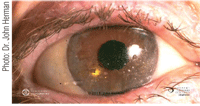
Depending on the patient’s ocular condition, he or she may wear their therapeutic lenses for a period of days to years. They may be utilized for daily or extended wear (see table 2). Because there is generally an underlying disease process precipitating the need for a therapeutic lens, extra caution must be taken to clean and disinfect the lens after wear, keeping in mind that silicone hydrogel lenses tend to deposit lipids more readily than HEMA lenses (see image 1). That said, the addition of a digital rubbing step is necessary for lenses that are used more than once.

It is critical to perform frequent follow-ups for bandage contact lens patients. One reason is that a bandage lens fit, by design, demonstrates less movement than a traditional soft lens fit. The theory is to provide increased patient comfort while preventing the healing epithelial cells from sloughing off due to any mechanical trauma of the lens itself.3 In addition, it is important for the practitioner to be vigilant regarding the detection of signs of microbial keratitis. The compromised cornea—especially when wearing lenses in an extended wear modality—is at particular risk for infection.4

Indications
Bandage contact lenses are indicated for many different reasons, including: protecting the eyes, increasing comfort, facilitating healing and sealing wound leaks. We’ll explore these indications, and others, in more detail in the following paragraphs.

• Protection. Corneal protection is needed in the case of several conditions, including: entropion, trichiasis, tarsal scars, recurrent corneal erosion, post-surgical ptosis and surgical sutures or exposed suture knots.

Recurrent erosions are a typical sequella of epithelial basement membrane (basal lamina) trauma or are secondary to anterior basement membrane dystrophy, anterior basement membrane degeneration or stromal dystrophy. A bandage contact lens is the second line of treatment, after hyperosmotic drops and/or ointment fail.2,5 An added benefit is the enhanced vision provided by the smooth refracting surface of the contact lens, as opposed to an irregular anterior corneal surface.5 Hypertonic saline drops should continue to be utilized concurrently with the lenses.

• Pain relief. The mitigation of corneal pain is another important indication for therapeutic contact lenses. The conditions most in need of this therapy include bullous keratopathy; epithelial erosion and abrasion; filamentary keratitis; and postoperative penetrating keratoplasty.

In bullous keratopathy, endothelial failure results in corneal edema, which in turn creates epithelial blisters that rupture, causing pain, foreign body sensation, and photophobia. A bandage contact lens reinforces the damaged tissues and protects the nerve endings from the abrasive actions of the eyelids. Patients who are awaiting a conjunctival flap or cornea transplant may be fitted with therapeutic lenses for up to 30 days at a time.2
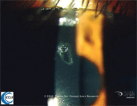
Until recently, pressure patching was the standard of care for treating large epithelial abrasions (see figure 2). With this treatment, the caveat was to refrain from patching contact lens wearers or injuries caused by presumed vegetative matter or false fingernails. The utilization of a bandage contact lens provides protection and healing for all three of these conditions, and it has now supplanted patching as the standard of care. The authors of the Wills Eye Manual caution, however, that prophylactic topical antibiotics should be used concurrently and that daily follow-up care is mandatory.6

1. Therapeutic soft contact lens with 2+ surface coating.

The other advantage of bandage contact lenses over patching is the ability to continue to install topical ophthalmic medications. This is particularly important after a corneal abrasion, erosion, or corneal refractive surgery, which necessitate the frequent installation of antibiotics and/or artificial tears. Some reports caution against the installation of cycloplegic agents (which reduce the pain associated with a corneal abrasion/erosion or after corneal refractive surgery) in bandage CL wearers. The dilating drops can cause the bandage lenses to dry out and become less comfortable, especially overnight, with the end result a potentially decreased healing response.7 On the other hand, bandage contact lenses can be utilized—by design—as vehicles for drug delivery, but the exact way to ensure a consistent dosage is still under investigation.

• Healing. The use of bandage contact lenses to facilitate healing is particularly necessary for the following conditions: chronic epithelial defects, corneal ulcer, neurotrophic keratitis, neuroparalytic keratitis, chemical burns and basement membrane disease.

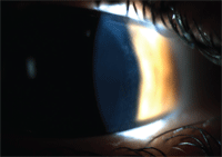
They also enhance healing following corneal surgery, particularly refractive surgery. They protect the cornea from exposure or from the irritation caused by rubbing the eye as the corneal wounds are healing. Therapeutic bandage contact lenses are a mainstay after photorefractive keratectomy (PRK) procedures, in which the removal of the epithelium leaves an open wound that takes about one week to heal (see figure 3). They are also valuable for the following procedures: laser-assisted in situ keratomileusis (LASIK), laser-assisted subepithelial keratomileusis (LASEK), Epi-LASIK, penetrating keratoplasty (PK) and phototherapeutic keratectomy (PTK), lamellar grafts and corneal flaps.

2. The use of therapeutic contact lenses has replaced pressure patching as the standard of care for epithelial abrasions such as the one in the image above.

• Sealing. The lenses also may aid in sealing leaky wounds. Serving as a splint or sealant, the lenses can be beneficial after cataract, penetrating keratoplasty or glaucoma filtering surgery.

• Maintenance of corneal hydration. The role of bandage contact lenses in dry eye is controversial. For patients who need to continually instill lubricating drops into their eyes, particularly after refractive surgery, the benefits of using a bandage lens can be great. Other patients who benefit are those who have significant lagophthalmos and subsequent corneal exposure. However, contact lenses are generally contraindicated for dry eye.7

• Structural stability and protection in piggyback lens fitting. Many patients benefit from the utilization of a soft and rigid lens concurrently. The rigid lens provides crisp vision, particularly for irregular corneas, and the soft bandage lens protects the cornea, preventing irritation and abrasions. Examples include elevation differences in the host/graft junction, keratoconus and in the presence of scar tissue.

Contraindications

3. The cornea, one day following PRK.

Each clinician must assess his patient’s condition carefully to determine whether a bandage contact lens is warranted. Interestingly, many of the conditions that require bandage contact lenses (dry eye, infection, inflammation, etc.) contraindicate lens wear in general. In addition, therapeutic contact lenses should not be used in patients who are unwilling or unable to comply with the necessary treatment and follow-up. 

Dr. Gromacki is a diplomate in the Cornea, Contact Lens and Refractive Technologies Section of the American Academy of Optometry. She is Chief Research Optometrist at Keller Army Community Hospital, West Point, New York.

1. Arrington GE. A history of ophthalmology. MD Publishers, New York, New York, 1959.
2. Weiner BM. Therapeutic bandage lenses. In: Silbert JA, ed. Anterior Segment Complications of Contact Lens Wear. Churchill Livingstone, New York, New York, 1994; 455-471.
3. Aquavella JV. Chronic corneal edema. Am J Ophthalmol 1973;(76):201-207.
4. Thoft RA, Mobilia EF. Complications with therapeutic extended wear soft contact lenses. Int Ophthalmol Clin. 1981;(21):197.
5. Chan WE, Weissman BA. Therapeutic contact lenses. In: Bennett ES, Weismann BA, (eds). Clinical Contact Lens Practice, Lippincott Williams & Wilkins: New York, New York, 2005: 619-628.
6. Ehlers JP and Shah CP, eds. The Wills Eye Manual Fifth Edition, Wolters Kluwer Health, New York, New York, 2008:15-16.
7. Russell GE. Bandage lenses: new opportunities in practice. Contact Lens Spectrum. 2004(6).
8. Tyler Thompson TT. Tyler’s Quarterly 2011;28(3):32-52.
9. White P. 2011 Contact Lenses and Solutions Summary. In: Contact Lens Spectrum (suppl) 2011;27(7):14.

Glare Reducing Lenses: Understanding Their Uses

Glare Reducing Lenses: Understanding Their Uses

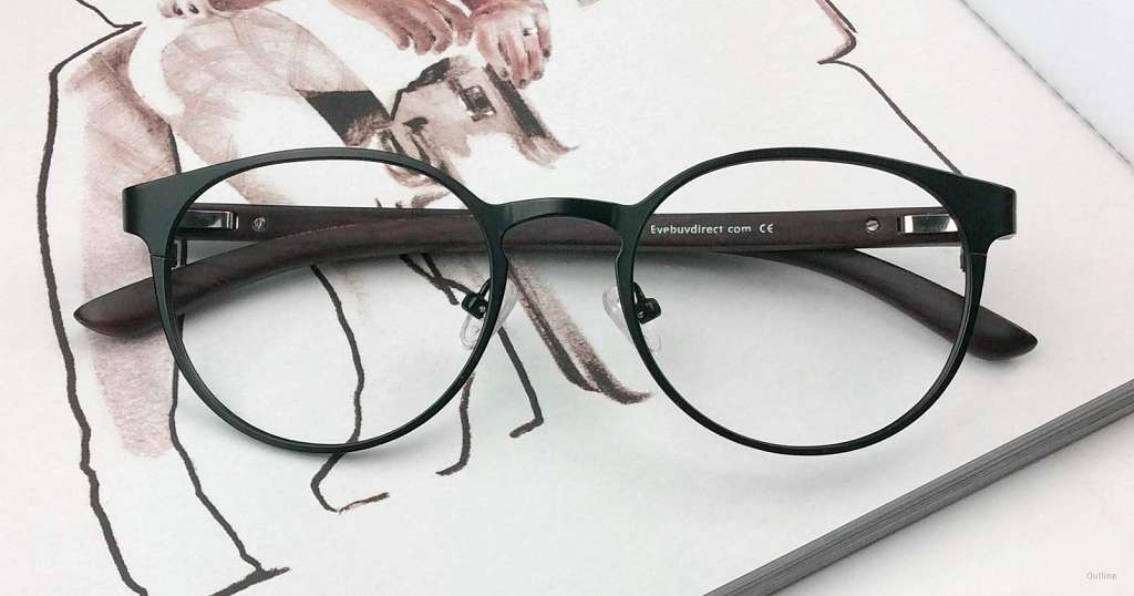
There are two main types of glare reducing lenses for eyewear: lenses with an anti-reflective coating and polarized lenses. Both help to prevent glare in their own way. Glare reducing lenses can improve vision clarity, help people see better while driving at night, reduce annoying glare from water or other horizontal surfaces, and eliminate noticeable reflections on a lens itself.

What is glare?

Glare is caused by light bouncing off of a reflective surface. When talking about eyewear, people are most likely referring to lens glare or environmental glare. Lens glare is caused by the reflection of light off the surface of a lens. Whether it belongs to a camera, telescope, binoculars, or even just glasses, all lenses have some level of reflection with the lowest amount of reflection being less than 0.1%. Eyeglass lenses without a glare-reducing coating typically allow around 90% of light to pass through, depending on the lens material. The other 10% of the light reflects off the surfaces of the lens. The glare caused by this 10% reduces vision clarity, causes people to see halos around headlights and street lamps at night, and creates bright, almost white reflections on the lens itself.

Environmental glare is caused by light waves reflecting off of flat surfaces like water or the highway. It becomes focused and travels in a uniform direction parallel to that surface, creating a bright and intense reflection that we call glare. This type of glare affects everyone, regardless of whether or not they wear glasses.

glare reducing lenses

Outline, in Black

How do glare reducing lenses work?

While it may be impossible to eliminate 100% of the glare on glasses lens, technology has helped to get the number as close to 0 as possible. While both anti-reflective coatings and polarized lenses help to reduce glare, the technology behind these two is quite different. An anti-reflective coating (also known as AR or anti-glare coating) actually encourages more light to pass through a lens. When more light passes through, less light reflected off its surfaces, and thus, less glare.

Polarized lenses, on the other hand, reduce glare by absorbing light waves from a certain orientation. Most polarized lenses for eyewear are oriented to absorb horizontal light waves reflected off of flat surfaces like a lake or the snow-covered ground.

When it comes to eyewear, AR coatings are applied both eyeglass lenses and sunglass lenses. Anti-glare coating is applied to both sides of a lens to prevent light from reflecting off the back of the lens as well. Polarized lenses are typically used for sunglasses since the nature of its glare reducing technology is to block light instead of letting more through.

The benefits of glare reducing lenses

Many people question whether or not it’s worth it to get glare reducing lenses. The short answer is: while not everyone may need sunglasses with polarized lenses, lenses with an anti-reflective coating will vastly improve the quality of life for a glasses wearer.

Lens glare is a major source of eye strain since it reduces vision clarity, forcing your eyes to work harder to focus. People who work with computers are especially susceptible to this type of eye strain since illuminated screens act as a direct and constant source of glare on lenses. Adding AR coating to your lenses significantly lessens this glare, helps you see more clearly, and reduces eye strain caused by computer screens.

Glares called “halos” can be seen around the headlights of cars and street lamps. These halosare a great source of discomfort and distraction for glasses wearers who drive at night. They reduce visibility and make nighttime driving difficult. Anti-glare coating prevents these halos and helps to make driving at night safer for glasses wearers.

If you’re someone who is both literally and figuratively in the spotlight a lot, anti-reflective coating is a must. Glare caused by bright lights reflecting off a lens can be distracting. It also obscures your eyes, making it harder for people to find direct eye contact with you. So if you have a client- or customer-facing job, make sure to consider getting glare reducing glasses.

Finally, if you’re someone who spends a lot of time out on the water or working in the snow, you’re well aware of how much glare can reduce visibility and make it a literal pain to be outside. The tint on sunglasses with polarized lenses helps to reduce that all around brightness, while the polarization helps to save your eyes from blinding glare.

So for the best comfort while wearing glasses or sunglasses, consider glare reducing lenses. Not only will they help you see better, but they’ll also help you get the most out of life. At EyeBuyDirect, you can find affordable glare reducing lenses for any of our great styles.

Coined from eyebuydirect.com

Buy infrared thermometer in Omole Ojodu Berger Magodo

You can buy 3 in 1 smart infrared thermometers at Eyeupdate clinic & optical supplies located at 01 Ajuwon junction, Ajuwon bus stop, beside BNPL filling station, Ajuwon near the grailand estate gate.

This is a non contact thermometer which can be used to take temperature measurement of the human body near the forehead, ear or wrist. Other features of the thermometer are:

Fast measurement (1 second)
Fever warning
Measurement range: 32. 0°C – 42. 2°C
Memory recall: 32 readings
Automatic shutdown: 60s
Error Resolution: 0.1°C
Can measure body temperature on human forehead
Can measure body temperature on the wrist
Can measure human temperature on the ear

Manufacturers: Alicn Medical Inc

Price: N35, 000
Tel: 08107531046
Address : Eyeupdate clinic & optical supplies,
01, Ajuwon junction, Ajuwon bus stop, off Elliott bus stop, Iju-Ishagah (beside BPNL Filling station, Ajuwon)

Buy infrared thermometer in Alagbole Akute Ajuwon Matogun

Eye clinics in Ojodu Berger Omole

Edited By: Dan Gudgel
Mar. 27, 2020

It is possible to do some basic testing of your vision and the vision of your family members and friends at home. Home eye testing is not a substitute for a complete medical eye examination by an ophthalmologist. Testing your vision at home will not be as accurate as what your ophthalmologist can do. But home eye testing could help you discover a problem that requires professional attention.

Many eye disorders can be corrected if discovered and treated early. Learn more about when eye exams are recommended.

Children under age 3 should have their vision tested by an optometrist, ophthalmologist or other vision care professional.

What You Need to Test Your Vision at Home

  • Something to cover the eye, like a paper cup or facial tissue.
  • Scissors.
  • Tape or tack to hang the test chart on the wall.
  • A pencil or pen to record the results.
  • A yardstick, tape measure, or ruler.
  • A flashlight, if available.
  • A well-lighted room at least 10 feet long.
  • The correct testing chart.

Prepare the Test Area

  • Select either the child’s or adult’s test chart and print it out.
    • When printed, the largest letter at the top of the chart should be just under an inch (23 millimeters) tall.
  • Measure 10 feet from a wall with no windows, and place a chair at this point.
  • Tape or pin the chart on the bare wall, level with the eyes of the person you will test as he or she sits in the chair.

Testing a Child (Age 3 or Older)

Explain to your child that you are going to play a “pointing game.” Using the practice E card, show him or her how to point in the same direction that the E is “pointing.” Turn the practice E in the four different directions (up, down, right, left). You may hold the practice card as close as the child wants until he or she can point in the four directions without help.

  • Have your child sit in the chair 10 feet from the chart, holding the cover over one eye without applying any pressure. Do not let the child peek. A second person may be needed to hold the cover in place and watch for peeking. If your child wears glasses, he or she should wear them during the test.
  • If the chart seems too dark to see clearly, use the flashlight to illuminate the test letters.
  • Point at each of the Es, starting with the largest. Have your child point in the direction the E is pointing.
  • Write down the number of the smallest line your child can correctly see (more than half of the Es correctly identified).
  • Then repeat the test with the other eye covered. If your child is tired, you may wish to test the other eye at a different time.

Testing an Adult or Older Child

  • Have the person being tested sit in the chair, 10 feet from the chart. Make sure the chart is level with his or her eyes.
  • Have the person being tested cover one eye. If he or she uses eyeglasses for distance vision, the glasses should be worn during the test.
  • Shine the flashlight on each line of the chart, while the person you are testing reads the letters out loud. Continue to the bottom row or until the letters are too difficult for the person to see.
  • Write down the number of the smallest line seen correctly (the line with more than half of the letters correctly identified).
  • Now repeat the test with the other eye covered and record the results.

What Are Normal Scores for Home Eye Testing?

A child should be able to see the 20/40 line by age 3 or 4 and the 20/30 line by age 5. If you test your child several times on different days and your child cannot see the expected line of print or cannot see the same line with each eye, he or she may have an eye problem. You should have your child evaluated by a physician.

An older child or adult should be reading the 20/20 line. You should arrange for a medical eye examination by an ophthalmologist if there are abnormal results.

Home Vision Test Results

Record the results of your home screening by filling in the number of the smallest line the person could read for each line below. If the test results indicate that you or your child needs to see an ophthalmologist, take the numbers you wrote down with you.

right eye left eye
Home Visual Acuity Screening 20/___ 20/___

Download Home Eye Testing Charts