Four technologies that could revolutionize the treatment of blindness

by Simon Makin

A decade ago, clinicians had nothing to offer most people affected by retinal degeneration. Breakthroughs in genetics, bionics and stem-cell therapy are changing that.

Worldwide, 36 million people have total vision loss1. They cannot see shapes or even sources of light. For most of these people, their blindness stems from rectifiable problems such as cataracts — they simply lack access to appropriate health care. The remaining millions, however, are blind as a result of conditions that currently have no effective treatment

Blindness is one of the most life-altering conditions a person could experience,” says William Hauswirth, an ophthalmologist at the University of Florida in Gainesville. As well as the difficulties that it causes for mobility and in finding employment, visual impairment is associated with a host of other health issues, including insomnia, anxiety and depression, and even risk of suicide. “Restoring useful vision would make an almost unimaginable improvement in quality of life,” Hauswirth says.

In high-income countries where preventable causes of visual impairment are routinely addressed, the leading cause of blindness is degeneration of the retina. Found at the back of the eye, this tissue contains specialized cells that react to light and process visual signals, and is therefore crucial to vision. Photoreceptor cells — neurons commonly known as rods and cones — convert light that strikes the retina into electrochemical signals. These signals then filter through a complex network of other neurons, including bipolar cells, amacrine cells and horizontal cells, before reaching neurons known as retinal ganglion cells. The long projections, or axons, of those cells form the optic nerve, along which signals from the retina are carried to the brain’s visual cortex, where they are interpreted as images.

Retinal disorders commonly involve the loss of photoreceptor cells, which depletes the eye’s sensitivity to light. In some retinal disorders, including age-related macular degeneration (AMD), this loss results from the failure of the epithelial cells that form a layer at the back of the retina known as the retinal pigment epithelium (RPE). The RPE keeps photoreceptor cells healthy by cleaning up toxic by-products produced during the reaction with light, as well as by providing nutrients. In retinal disorders in which photoreceptors remain in good shape, the main cause of blindness is degeneration of retinal ganglion cells.

Variety in the causes of visual impairment makes it more difficult to find solutions. But advances in several areas are raising hopes that almost all forms of retinal disorder could become treatable.

One approach is to augment or bypass damaged eyes with functional prostheses. Such bionic eyes can restore only limited vision at present, but researchers continue to push the devices’ capabilities. Another option is gene therapy. Already available to people with specific genetic mutations, researchers are looking to extend this approach to more people and conditions. Some scientists are also pursuing treatments based on a related technique known as optogenetics, which involves genetically altering cells to restore light sensitivity to the retina. This work is at an early stage, but researchers hope that the approach will ultimately be able to help a wide range of people, because it is agnostic to the causes of retinal degeneration. And efforts to replace lost or damaged cells of the retina, either in situ or through cell transplants, hint that even late-stage retinal disorders might eventually become treatable.

Much of this research is in its infancy. But Hauswirth is upbeat about the progress that has already been made. Ten years ago, he says, he often had to tell patients that he could do nothing for them. “For many of these diseases, that’s totally changed.”

Bionic eyes

Almost 30 years ago, Mark Humayun, a biomedical engineer at the University of Southern California in Los Angeles, began to electrically stimulate the retinas of people with blindness. Working with colleagues at Second Sight Medical Products, a medical technology firm in Sylmar, California, his experiments showed that such stimulation could induce the visual perception of spots of light called phosphenes. After a decade of work in animals to establish the amount of electrical current that could be applied safely to the eye, and armed with vastly increased knowledge about the number and types of cell that persist in degenerating human retinas, Humayun’s team was ready to begin working with people. Between 2002 and 2004, the researchers implanted a bionic eye in each of six people who had total or almost-total blindness in one eye — the first trial of its kind. Recipients of the device, known as the Argus I, reported being able to perceive phosphenes, directional movement and even shapes2. Around 300 people now experience the world through that device’s successor, the Argus II, which was approved by regulators in Europe in 2011 for use in people with retinitis pigmentosa — a group of rare genetic disorders that cause photoreceptor cells to degenerate. The US Food and Drug Administration (FDA) followed suit two years later.

To be fitted with an Argus II, patients undergo surgery to attach a chip containing an electrode array to the surface of the retina. To ‘see’ with the device, a miniature video camera mounted on a pair of glasses relays signals to a processing unit that is worn by the recipient. The processor converts the signals into instructions that are transmitted wirelessly to the implanted device. The electrodes then stimulate retinal ganglion cells at the front of the retina. Using the prosthesis is a learning process. Recipients must train their brain to interpret the new type of information being received. And because the video camera does not track the motion of the eye, they must also learn to move their head to direct their gaze.

The device provides only limited vision. Users can detect light sources and objects with high-contrast edges, such as doors or windows, and some can decipher large letters. These limitations arise partly because the device’s 60 electrodes provide very low resolution compared with the millions of photoreceptor cells in a healthy eye. But even this minimal enhancement can improve people’s lives considerably.

Whereas the Argus II is an epiretinal implant — meaning that it lies on the surface of the retina — other devices in development are designed be placed beneath the retina. These subretinal implants can stimulate cells that are closer to those that normally introduce signals to the retina — the photoreceptor cells. By stimulating cells higher up in the visual pathway, researchers hope to preserve more of the signal processing that is performed by a healthy retina.

Retina Implant, a biotechnology company based in Reutlingen, Germany, has built a subretinal implant comprising photodiodes (semi-conductor devices that convert light into electrical current) that directly sense light entering the eye. This eliminates the need for an external video camera, enabling users to direct their gaze naturally. Power is supplied by a hand-held unit, through a coil that is implanted under the skin above the ear. Alpha AMS, the current version of the system, has received regulatory approval in Europe for use in people with retinitis pigmentosa.

Pixium Vision in Paris is testing a photovoltaic subretinal implant called Prima. The system projects signals from a video camera mounted on glasses into the eye using near infrared light, the wavelength of which optimally drives photodiodes in the device to stimulate retinal cells. Projecting images in this way gives users some control over the direction of their gaze, because they can explore the scene by moving just their eyes. Power is also provided by the near infrared light, making the implant wireless and the surgery to fit it less complicated. “Patients are learning how to regain vision faster, and the resolution seems better,” says José-Alain Sahel, an ophthalmologist at the University of Pittsburgh, Pennsylvania, who is conducting safety trials of the device in ten people with AMD. “It’s early days, but this is very promising.”

All of these devices work only when functioning cells remain in the retina. In common eye conditions that affect mainly photoreceptor cells, including retinitis pigmentosa and AMD, there are usually some cells left to stimulate. But when too many retinal ganglion cells die, as occurs in advanced diabetic retinopathy and glaucoma, such implants cannot help. For people without any remaining retinal function, whether due to disease or injury, an alternative bionic approach might be more relevant.

Humayun and his colleagues are working on a system that bypasses the eye by sending signals straight to the brain. The idea is not new: in the 1970s, US biomedical engineer William Dobelle showed that directly stimulating the visual cortex triggered the perception of phosphenes3. But bionic-eye technology is only now catching up. Second Sight has developed Orion, a system that is, according to Humayun, “basically a modified Argus II”. Similarly to the original, it uses a video camera and signal processor that communicate wirelessly with an implant, but the chip is placed on the surface of the visual cortex rather than on the retina. The device is being tested in five people with limited or no light perception owing to an eye injury or damage to the retina or optic nerve. “So far, the results are good,” he says. “We’re not surprised by anything yet.”

Given that some of the technology is already tried and tested in people, Humayun is optimistic that the system could receive regulatory approval within a few years. “Obviously, brain surgery has a different level of risk, but the procedure is pretty straightforward, and the Orion could help a lot more patients,” he says. However, much less is known about stimulating the brain to provide useful vision. “We know a lot about the retina but very little about the cortex,” says Botond Roska, a neurobiologist at the Institute of Molecular and Clinical Ophthalmology Basel in Switzerland. “But we’ll never know enough if we don’t try,” he says.

Gene therapy

The eye is an ideal target for gene therapy. Because it is relatively self-contained, the viruses that are used to carry genes into the cells of the retina should not be able to travel to other parts of the body. And because the eye is an immunoprivileged site, the immune system is less likely to mount a defence there against such a virus.

In the first demonstration of gene therapy’s potential for tackling blindness, three teams of researchers have used the technique to successfully treat people with Leber congenital amaurosis (LCA). This inherited condition leads to severe visual impairment and begins in the first few years of life, often manifesting as night blindness before progressing to broad vision loss that starts at the periphery of the visual field. It affects about 1 in 40,000 babies.

The researchers set out to tackle a specific form of the condition known as LCA 2. This is caused by mutations in RPE65, a gene that is expressed by the RPE. The mutated gene adversely affects RPE function, which in turn damages photoreceptor cells. In 2008, the three teams, including one led by Hauswirth, each showed in early-stage clinical trials that delivering a healthy copy of RPE65 to the retina was safe and led to limited improvements in vision4,5,6. A phase III clinical trial led by Albert Maguire, an ophthalmologist at the University of Pennsylvania in Philadelphia, showed in August 2017 that people with LCA 2 who received the treatment were better able to navigate obstacle courses at various levels of illumination than those who did not7. In December 2017, the FDA approved the treatment, voretigene neparvovec (Luxturna), making it the first gene therapy for any condition to get the green light for clinical use.

It is possible to treat LCA 2 in this way because the genetic mutations involved show a recessive pattern of inheritance. This means that both of a person’s copies of RPE65 must carry the relevant mutations to cause the disorder. Supplying a single, unmutated version therefore fixes the problem. Conditions that are caused by dominantly inherited mutations, however, require only one mutated copy of a gene to manifest. In most of these, simply adding a normal copy of the gene will not help; instead, the mutated gene must be inactivated. One option is to silence it by adding specific RNA molecules that intercept the mutated gene’s instructions for making the faulty protein, and then supplying a normal copy of the gene to take over its duties — an approach termed suppression and replacement. Another is to correct the mutation using the gene-editing technique CRISPR–Cas9. Researchers at the University of Modena and Reggio Emilia in Modena, Italy, demonstrated this approach in a mouse model of retinitis pigmentosa8 in 2016. The following year, a team in the United States used it to correct the mutation that causes a type of glaucoma both in mice and in cultured human cells9.

An important driver of gene therapy’s progress has been the use of adeno-associated virus (AAV) to deliver replacement genes to cells. AAVs have been shown to be safe, in part, because they tend not to integrate into their host cell’s genome, which minimises the risk of cells turning cancerous. And their small size enables them to diffuse widely through the eye and therefore infect a large number of cells. But the ability of AAVs to deliver genes has limits: some genes are simply too large for AAVs to carry, including ABCA4, mutations in which can lead to Stargardt disease, an inherited form of macular degeneration. Two workarounds are being pursued. The first uses a virus with a greater carrying capacity, such as a lentivirus, to deliver replacement genes. The safety and efficacy of this approach is unknown but clinical trials are under way. A second strategy is to break the replacement gene in two and transport each half separately into the cell, together with a means of recombining them. “That’s working in at least one animal model right now,” says Hauswirth.

Regardless of the approach, gene therapy has a considerable limitation. More than 250 genes are implicated in blindness, and because each can be affected by numerous types of mutation, the number of potential therapeutic targets is enormous. For example, more than 100 mutations in the gene RHO lead to retinitis pigmentosa, the most common dominantly inherited retinal disorder. Developing a gene therapy for each and every mutation is not practical, says Hauswirth.

Researchers are working on a potential solution that puts a twist on the suppression-and-replacement approach. Instead of targeting copies of RHO containing a specific mutation, they use a silencing RNA to suppress all expression of the gene, whether RHO is mutated or not, while delivering a replacement copy that is immune to the silencing RNA. A team led by Jane Farrar, a geneticist at Trinity College, Dublin, showed the promise of this strategy in 2011 in a mouse model of dominant retinitis pigmentosa10. In 2018, Hauswirth and colleagues tested the approach in dogs with retinitis pigmentosa11. They showed that degeneration of photoreceptor cells in treated areas of the retina could be halted — an improvement that persisted for at least eight months. This strategy tackles all mutations that can cause dominantly inherited retinitis pigmentosa in a single treatment, and therefore extends gene therapy from recessive to dominantly inherited conditions “in a fairly simple way”, Hauswirth says. He plans to study how well dogs that have received the treatment can navigate a maze, and is collecting the safety data required to start a clinical trial.

Optogenetics

Gene therapy works only in people whose blindness is caused by genetic mutation. It is also not appropriate for tackling end-stage retinal disease, in which an insufficient number of cells remain to be repaired. But a related approach based on a technique called optogenetics is disorder agnostic and could lead to treatments for different stages of degeneration. In optogenetics, genes that enable cells to produce light-sensitive proteins known as opsins are delivered by a virus. Introducing opsins can restore some light sensitivity to damaged photoreceptors, or even make other cells of the retina, including bipolar cells or retinal ganglion cells, sensitive to light.

Problematically, however, whereas photoreceptor cells in the eye can cope with a wide range of light intensities — working well in both bright sunlight and twilight — opsins have a limited range and often perform better at high light intensities. A potential solution is to use a set-up that works in a similar way to Pixium Vision’s Prima bionic-eye system, in which recipients are fitted with glasses that incorporate a video camera that captures the user’s view and a projector that points into their eye. As with Prima, the benefit is that the nature of the light that enters the eye can be tailored to the retina’s modification; however, in this case, the intensity and wavelength chosen are those that best drive the newly introduced opsins rather than implanted photodiodes.

GenSight Biologics, a biotechnology company in Paris that counts Sahel and Roska among its founders, is already testing such a system. It aims to deliver an opsin to retinal ganglion cells, but there is a potential snag: retinal ganglion cells are naturally sensitive to light. They express melanopsin, a protein involved in the pupillary light reflex, in which the pupil of the eye constricts in response to bright light. To avoid triggering this, the researchers at GenSight are using an opsin that responds to red wavelengths of light, because melanopsin responds preferentially to light at the blue end of the spectrum. The company began an early-stage clinical trial in October 2018 in people with advanced retinitis pigmentosa who have minimal sight remaining. The trial will involve cohorts from the United Kingdom, France and the United States, and the initial results are expected by the end of 2020.

“This is a simple approach, and we’ll have to see what will be gained,” Roska says. “Then, we can move to more and more sophisticated approaches.” One problem that remains is that many of the disorders that optogenetic techniques might treat involve degeneration of specific parts of the retina, with useful vision being retained in other areas. The light that drives opsins is visible and could interfere with remaining natural vision. In the future, opsins that respond to near infrared light might enable optogenetics treatments to work in tandem with residual natural vision.

Cell regeneration

Stem-cell therapy could potentially cure blindness even in the late stages of disease. Because stem cells can be coaxed into becoming any type of cell, they could be used to grow fresh retinal cells for transplantation into the eye to replace those that have been lost. However, studies in animals have shown that only a small proportion of transplanted neurons are able to integrate correctly into the retina’s complex neural circuitry. This is a considerable obstacle for stem-cell treatments that aim to replace retinal neurons.

The cells that make up the retinal pigment epithelium, on the other hand, sit outside the retina’s circuitry. Stem-cell-based therapies therefore hold most promise for conditions, such as AMD and retinitis pigmentosa, that cause RPE cells to degenerate. “Photoreceptors have to connect to the circuitry but the retinal pigment epithelium does not,” says Roska. “That’s where people are closest to making advances.” Initially, researchers tried injecting the retina with stem-cell-derived RPE cells in suspension, but too few stuck around where they were needed. Several teams now think that a better approach is to transplant RPE cells into the eye as a preformed sheet that is then held in position by a biocompatible scaffold. “The scaffold approach is a huge improvement, compared to suspension, for RPE cells,” says Sahel.

In March 2018, the London Project to Cure Blindness — a collaboration between University College London and Moorfields Eye Hospital in London — announced the findings of a phase I trial in which a sheet of RPE cells was implanted in the retinas of two people with wet AMD (a rare, serious form of AMD involving abnormal growth and leakage of blood vessels). Both recipients tolerated the procedure well and were able to read 21–29 more letters on a reading chart than before the treatment12. The following month, a team led by Humayun reported similar phase I results in five people with dry AMD, the more common form of the condition13. These initial results are full of promise. “This has led to a lot of excitement,” says Humayun. But the findings need to be confirmed by phase III trials in a greater number of participants, and Humayun cautions that the treatment might be many years away from use in the clinic, because no stem-cell therapy for a retinal disorder has yet made it through the approval process.

A related approach, still in the early stages of basic research, could fulfil the hope of replacing lost neurons, opening the door to treatments for a wide variety of eye diseases. In humans, mature neurons do not divide and therefore cannot regenerate, which makes restoring vision especially difficult. But the same is not true of all animals. Reptiles and certain fish can regenerate retinal neurons, and birds also exhibit some regenerative capacity. Thomas Reh, a neuroscientist at the University of Washington in Seattle, is trying to unlock this ability in humans. But rather than transplanting cells grown in the laboratory, Reh aims to coax cells that are already in the retina to differentiate into fresh neurons.

In 2001, Reh suggested that Müller glia — cells that provide structure to the retina and support its function — are the source of new neurons that had been observed in fish and birds14. He and his team then set about finding out whether Müller glia could be used to generate fresh neurons in mice. In 2015, they engineered mice to make Ascl1, a protein that is important for producing neurons in fish, and then damaged the animals’ retinas15. Their hope was that Ascl1 would provoke Müller glia to transform into neurons.

The experiment failed to produce new neurons in adult mice, but succeeded in young mice. Nikolas Jorstad, a biochemist and PhD student in Rehs’ team, proposed that chemical modifications made to chromatin (a complex of DNA, RNA and proteins) in the cell nucleus during development might block access in mature cells to genes that enable Müller glia to transform into neurons. In August 2017, Reh’s team showed that by introducing an enzyme that reverses such modifications, they could coax Müller glia to differentiate16. “For the first time, we could regenerate neurons in the adult mouse,” Reh says. “After all these years I was pretty thrilled.” Although they were not true photoreceptor cells, and looked more like bipolar cells, the neurons connected to the existing circuitry, and were sensitive to light. “I was surprised they connect as well as they do,” says Reh.More from Nature Outlooks

Although far from being ready to treat retinal disorders in people, the work has huge potential. The next step will be to repeat the studies in animals with eyes that are more similar to those of humans. Reh’s team are already working with retinal cell cultures from non-human primates. The researchers also need to work out how to direct the differentiation process to produce specific cell types such as rods and cones. “Now we’ve got our foot in the neuron-making business, cones would be great,” says Reh.

If successful, the approach could be widely applicable. “Ultimately, this will be the way all these eye diseases will be treated,” Reh predicts. “It just makes sense. You don’t have to worry about getting transplants right. Your cells are right where you need them.”

Humayun is also encouraged by the work. “I cheer on anybody with a new good idea,” he says. “It’s very early, it’s high risk, but never say never. That’s what I’ve learned.”

References

  1. 1.Bourne, R. R. A. et al. Lancet Glob. Health 5, e888–e897 (2017).
  2. 2.Humayun, M. S. et al. Vision Res. 43, 2573–2581 (2003).
  3. 3.Dobelle, W. H. & Mladejovsky, M.

Eyeupdate Eye Clinic at 01, Ajuwon junction, Ajuwon bus stoo, Akute/Ajuwon Road, beside BPNL Filling Station, near Lambe Ogun State Tel: 08107531046, 07030000001, 08107531046.

This eye clinic is serving :

alagbole, akute, ajuwon, lambe, matogun, matogbun, giwa, okearo, koye, oke, arifanla, baale, Road, fagba, abule, egba, agege, ogba, ikeja, G. R. A, oshodi, Berger, ojodu, omole, magodo, yaba, mowe, ibafo, shagamu, Ishagah, shomolu, heritage, estate, ajao, College Road, iju, Road, water, works, omole, magado, ketu, ojota, ikoyi, akiode bus stop, grammar school, first gate, Ala Hausa, Yoruba, Igbo, Benin, Edo, Association, Club, Ikorodu, Ketu, Ojota, Shomolu, Cele, Ogun, Lagos, Lagos Island, Lagos mainland, Ifako, Ijaye, eti osa, Oyo, Lisa, Obawole,Iju, Ishagah, Ishaga, Isaga, Isagah, Ishasi,Obawale,Owode, sang ota, abule egba, redeem camp, Ijebu ode, toll gate, bus stop, Street, along, Nigeria

Key words related to eyecare:

Buy,optical, ophthalmic, instruments, hospital, instruments, beds, auto-refractors, keratometer, direct, ophthalmoscope, retinoscopes, optoscopes, lensmeter, slit-lamp, bio microscope, trial, lens, boxes, infrared thermometer, blood pressure monitor, acucheck, phoroptor, phoropter, chair unit, glazing, edging, machine, tonometer, optimal system,colour contact lenses, designer, frames, pupilometer, ventilators,

Gene therapy works only in people whose blindness is caused by genetic mutation. It is also not

Russian Covid-19 Vaccine very effective – British Medical Journal

According to RT Respected British medical journal The Lancet publishes study showing Russia’s ‘Sputnik V’ Covid-19 vaccine to be 100% effective

Respected British medical journal The Lancet publishes study showing Russia’s ‘Sputnik V’ Covid-19 vaccine to be 100% effective

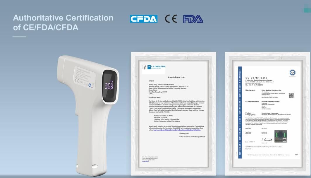
Buy non contact infrared thermometer within Akute, Ajuwon, Alagbole

https://opticalshop.eyeupdateclinic.com/product/buy-infrared-thermometer/

The world’s first registered Covid-19 vaccine successfully produced antibodies in all 76 participants in early-stage trials, according to a study published in The Lancet, one of the oldest and best-respected medical journals.

The trials of ‘Sputnik V,’ funded by the Russian Ministry of Health, discovered that every single patient who received the vaccine developed antibodies, and none showed any significant side effects.

On August 11, Russian President Vladimir Putin announced that the country had registered the world’s first Covid-19 vaccine. Developed by Moscow’s Gamaleya Research Institute of Epidemiology and Microbiology, the formula will first be distributed to teachers and medical workers before being made available to the general public next year.

Following its registration, scientists and epidemiologists worldwide criticized Russia for the vaccine’s rapid development, questioning its safety due to the small number of trial subjects. Although the testing was successful, longer-term trials, including a placebo comparison, are required to establish its actual quality, according to The Lancet.

However, according to the Russian Direct Investment Fund (RDIF), the scientific data provided in the article proves the “safety and effectiveness of the Russian vaccine.”

Explaining why it took a month to publish the results, Gamaleya Institute head, Alexander Gintsburg, told Russian news agency Interfax that it took a long time to prepare, and the article was evaluated by five independent reviewers, following all standard international peer-review conventions.

“The scientific community has assessed it quite objectively,” he explained

Despite its well-earned reputation, The Lancet has not been immune from controversy. Earlier this year, the journal published a study refuting the effectiveness of malaria drug hydroxychloroquine against Covid-19, but it was later withdrawn due to multiple errors.

Russia is not the only country racing to develop an effective vaccine against coronavirus. In July, a coronavirus vaccine developed by the University of Oxford was reported as creating immunity in a trial of 1,077 people. Earlier that month, UK security minister James Brokenshire claimed that Russian hackers had attacked British labs to steal vaccine research data.

Eye care tips

Your eyes are an important part of your health. Most people rely on their eyes to see and make sense of the world around them. But some eye diseases can lead to vision loss, so it is important to identify and treat eye diseases as early as possible. You should get your eyes checked as often as your health care provider recommends it, or if you have any new vision problems. And just as it is important to keep your body healthy, you also need to keep your eyes healthy.

Eye Care Tips

There are things you can do to help keep your eyes healthy and make sure you are seeing your best:

  • Eat a healthy, balanced diet. Your diet should include plenty or fruits and vegetables, especially deep yellow and green leafy vegetables. Eating fish high in omega-3 fatty acids, such as salmon, tuna, and halibut can also help your eyes.
  • Maintain a healthy weight. Being overweight or having obesity increases your risk of developing diabetes. Having diabetes puts you at higher risk of getting diabetic retinopathy or glaucoma.
  • Get regular exercise. Exercise may help to prevent or control diabetes, high blood pressure, and high cholesterol. These diseases can lead to some eye or vision problems. So if you exercise regularly, you can lower your risk of getting these eye and vision problems.
  • Wear Transition lenses. Sun exposure can damage your eyes and raise your risk of cataracts and age-related macular degeneration. Protect your eyes by using sunglasses that block out 99 to 100 percent of both UV-A and UV-B radiation.
  • Wear protective eye wear. To prevent eye injuries, you need eye protection when playing certain sports, working in jobs such as factory work and construction, and doing repairs or projects in your home.
  • Avoid smoking. Smoking increases the risk of developing age-related eye diseases such as macular degeneration and cataracts and can damage the optic nerve.
  • Know your family medical history. Some eye diseases are inherited, so it is important to find out whether anyone in your family has had them. This can help you determine if you are at higher risk of developing an eye disease.
  • Know your other risk factors. As you get older, you are at higher risk of developing age-related eye diseases and conditions. It is important to know you risk factors because you may be able to lower your risk by changing some behaviors.
  • If you wear contacts, take steps to prevent eye infections. Wash your hands well before you put in or take out your contact lenses. Also follow the instructions on how to properly clean them, and replace them when needed.
  • Give your eyes a rest. If you spend a lot of time using a computer, you can forget to blink your eyes and your eyes can get tired. To reduce eyestrain, try the 20-20-20 rule: Every 20 minutes, look away about 20 feet in front of you for 20 seconds.

Eye Tests and Exams

Everyone needs to have their eyesight tested to check for vision and eye problems. Children usually have vision screening in school or at their health care provider’s office during a checkup. Adults may also get vision screenings during their checkups. But many adults need more than a vision screening. They need a comprehensive dilated eye exam.

Getting comprehensive dilated eye exams is especially important because some eye diseases may not have warning signs. The exams are the only way to detect these diseases in their early stages, when they are easier to treat.

The exam includes several tests:

  • A visual field test to measure your side (peripheral) vision. A loss of peripheral vision may be a sign of glaucoma.
  • A visual acuity test, where you read an eye chart about 20 feet away, to check on how well you see at various distances
  • Tonometry, which measures your eye’s interior pressure. It helps to detect glaucoma.
  • Dilation, which involves getting eye drops that dilate (widen) your pupils. This allows more light to enter the eye. Your eye care provider examines your eyes using a special magnifying lens. This provides a clear view of important tissues at the back of your eye, including the retina, macula, and optic nerve.

If you have a refractive error and are going to need glasses or contacts, then you will also have a refraction test. When you have this test, you look through a device that has lenses of different strengths to help your eye care professional figure out which lenses will give you the clearest vision.

At what age you should start getting these exams and how often you need them depends on many factors. They include your age, race, and overall health. For example, if you are African American, you are at higher risk of glaucoma and you need to start getting the exams earlier. If you have diabetes, you should get an exam every year. Check with your health care provider about if and when you need these exams

Buy eye care products

Schedule your testOptical eye clinic in alagbole akute ajuwon matogun olambe lambe koye oke iju ishaga fagba

Hire or be hired http://jobs.eyeupdateclinic.com

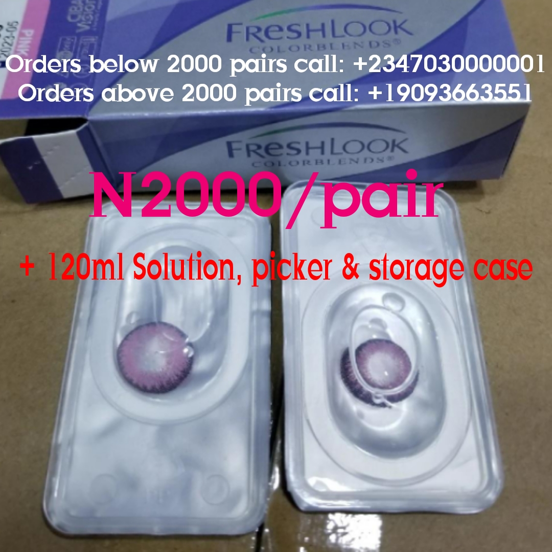
Buy freshlook contact lenses at N2000/pair with 120ml solution, storage case and picker within in Ajuwon Giwa Okearo Alagbole Akute

Buy Freshlook coloured contact lenses @ N2000/pair with 120ml solution and storage case and picker.
– Available quantity: 10, 000 pairs
– available colours: All freshlook colours…including all new unique colours
– Expiry Date: January 2024
– Made in USA
– Supply ability: 50, 000 pairs/ month
– Delivery coverage: Nigeria and West Africa
– Free pre-contact lens fitting examination and tutorial
– To order between 1 pair and 2000 pairs, call +2347030000001
– To order above 2000 pairs, call: +19093663551

COVID-19 & Eye Health -Eye/Optical within Obawole Iju Ishaga

The 2019 novel coronavirus (COVID-19) pandemic is rapidly changing the way the health and development communities are working. With the proximity of eye health professionals to patients during eye examinations and reports that the virus can cause conjunctivitis, COVID-19 has implications for eye health and eye health professionals. Travel restrictions, reprioritisation of health resources and economic consequences are impacting the work of many of our member and partner organisations.

To assist IAPB members, eye health personnel, health professionals and program personnel, IAPB is collating and sharing information and resources specific to eye health and international development in relation to COVID-19. Without much ado, COVID-19 Resources: Here is what we know:

COVID 19 & Eye Health Resources
COVID 19 Resources
RESOURCES 

Eye Health, Health, International Development and Vision Impairment News and Blogs

Member Stories
MEMBER STORIES

Read the COVID 19 stories from our Members across the globe.

Contact lenses -Eye/Optical clinic near Obawole Fagba Iju Ishaga

Contact lenses are an excellent choice for nearly anyone who needs vision correction and doesn’t want to wear eyeglasses full time or undergo LASIK surgery.

Unsure about contact lenses? This article will detail contact lens materials, contact lens designs and features and even new contact lens formats.

For instance, the first light-adaptive contact lenses, Acuvue Oasys with Transitions, debuted in the United States in 2019 and contact lenses embedded with antibiotics are in the works. (See our contact lens news page for the latest in contact lenses.)

Here are the basics you should know about contact lenses before seeing your eye doctor if you are interested in wearing contacts.

Contact Lens Materials

The first choice when considering contact lenses is which lens material will best satisfy your needs. There are five types of contact lenses, based on type of lens material they are made of:

ExpandableContact Lens Material

  • Soft lenses are made from gel-like, water-containing plastics called hydrogels. These lenses are very thin and pliable and conform to the front surface of the eye. Introduced in the early 1970s, hydrogel lenses made contact lens wear much more popular because they typically are immediately comfortable. The only alternative at the time was hard contact lenses made of PMMA plastic (see below). PMMA lenses typically took weeks to adapt to and many people couldn’t wear them successfully.
  • Silicone hydrogel lenses are an advanced type of soft contact lenses that are more porous than regular hydrogel lenses and allow even more oxygen to reach the cornea. Introduced in 2002, silicone hydrogel contact lenses are now the most popular lenses prescribed in the United States.
  • Gas permeable lenses — also called GP or RGP lenses — are rigid contact lenses that look and feel like PMMA lenses (see below) but are porous and allow oxygen to pass through them. Because they are permeable to oxygen, GP lenses can be fit closer to the eye than PMMA lenses, making them more comfortable than conventional hard lenses. Since their introduction in 1978, gas permeable contact lenses have essentially replaced nonporous PMMA contact lenses. GP contacts often provide sharper vision than soft and silicone hydrogel contacts — especially if you have astigmatism. It usually takes some time for your eyes to adjust to gas permeable lenses when you first start wearing them, but after this initial adaptation period, most people find GP lenses are as comfortable as hydrogel lenses.
  • Hybrid contact lenses are designed to provide wearing comfort that rivals soft or silicone hydrogel lenses, combined with the crystal-clear optics of gas permeable lenses. Hybrid lenses have a rigid gas permeable central zone, surrounded by a “skirt” of hydrogel or silicone hydrogel material. Despite these features, only a small percentage of people in the U.S. wear hybrid contact lenses, perhaps because these lenses are more difficult to fit and are more expensive to replace than soft and silicone hydrogel lenses.
  • PMMA lenses are made from a transparent rigid plastic material called polymethyl methacrylate (PMMA), which also is used as a substitute for glass in shatterproof windows and is sold under the trademarks Lucite, Perspex and Plexiglas. PMMA lenses have excellent optics, but they do not transmit oxygen to the eye and can be difficult to adapt to. These (now old-fashioned) “hard contacts” have virtually been replaced by GP lenses and are rarely prescribed today.

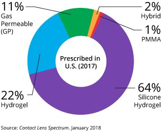
In 2017, 64 percent of contact lenses prescribed in the U.S. were silicone hydrogel lenses, followed by soft (hydrogel) lenses (22 percent), gas permeable lenses (11 percent), hybrid lenses (2 percent) and PMMA lenses (1 percent).


Home Contact Lenses Soft Contact Lenses |  En Español

Contact lens basics: Types of contact lenses and more

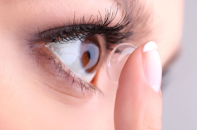
woman applying a contact lens

Contact lenses are an excellent choice for nearly anyone who needs vision correction and doesn’t want to wear eyeglasses full time or undergo LASIK surgery.

Unsure about contact lenses? This article will detail contact lens materials, contact lens designs and features and even new contact lens formats.

For instance, the first light-adaptive contact lenses, Acuvue Oasys with Transitions, debuted in the United States in 2019 and contact lenses embedded with antibiotics are in the works. (See our contact lens news page for the latest in contact lenses.)

Here are the basics you should know about contact lenses before seeing your eye doctor if you are interested in wearing contacts.

Contact Lens Materials

The first choice when considering contact lenses is which lens material will best satisfy your needs. There are five types of contact lenses, based on type of lens material they are made of:

ExpandableContact Lens Material

  • Soft lenses are made from gel-like, water-containing plastics called hydrogels. These lenses are very thin and pliable and conform to the front surface of the eye. Introduced in the early 1970s, hydrogel lenses made contact lens wear much more popular because they typically are immediately comfortable. The only alternative at the time was hard contact lenses made of PMMA plastic (see below). PMMA lenses typically took weeks to adapt to and many people couldn’t wear them successfully.
  • Silicone hydrogel lenses are an advanced type of soft contact lenses that are more porous than regular hydrogel lenses and allow even more oxygen to reach the cornea. Introduced in 2002, silicone hydrogel contact lenses are now the most popular lenses prescribed in the United States.
  • Gas permeable lenses — also called GP or RGP lenses — are rigid contact lenses that look and feel like PMMA lenses (see below) but are porous and allow oxygen to pass through them. Because they are permeable to oxygen, GP lenses can be fit closer to the eye than PMMA lenses, making them more comfortable than conventional hard lenses. Since their introduction in 1978, gas permeable contact lenses have essentially replaced nonporous PMMA contact lenses. GP contacts often provide sharper vision than soft and silicone hydrogel contacts — especially if you have astigmatism. It usually takes some time for your eyes to adjust to gas permeable lenses when you first start wearing them, but after this initial adaptation period, most people find GP lenses are as comfortable as hydrogel lenses.
  • Hybrid contact lenses are designed to provide wearing comfort that rivals soft or silicone hydrogel lenses, combined with the crystal-clear optics of gas permeable lenses. Hybrid lenses have a rigid gas permeable central zone, surrounded by a “skirt” of hydrogel or silicone hydrogel material. Despite these features, only a small percentage of people in the U.S. wear hybrid contact lenses, perhaps because these lenses are more difficult to fit and are more expensive to replace than soft and silicone hydrogel lenses.
  • PMMA lenses are made from a transparent rigid plastic material called polymethyl methacrylate (PMMA), which also is used as a substitute for glass in shatterproof windows and is sold under the trademarks Lucite, Perspex and Plexiglas. PMMA lenses have excellent optics, but they do not transmit oxygen to the eye and can be difficult to adapt to. These (now old-fashioned) “hard contacts” have virtually been replaced by GP lenses and are rarely prescribed today.

In 2017, 64 percent of contact lenses prescribed in the U.S. were silicone hydrogel lenses, followed by soft (hydrogel) lenses (22 percent), gas permeable lenses (11 percent), hybrid lenses (2 percent) and PMMA lenses (1 percent).

LEARN MORE about the proper care of your contact lenses.

Contact Lens Wearing Time

Until 1979, everyone who wore contact lenses removed and cleaned them nightly. The introduction of “extended wear” enabled wearers to sleep in their contacts. Now, two types of lenses are classified by wearing time:

  • Daily wear — must be removed nightly
  • Extended wear — can be worn overnight, usually for seven days consecutively without removal

“Continuous wear” is a term that’s sometimes used to describe 30 consecutive nights of lens wear — the maximum wearing time approved by the FDA for certain brands of extended wear lenses

When To Replace Your Contact Lenses

Even with proper care, contact lenses (especially soft contacts) should be replaced frequently to prevent the build-up of lens deposits and contamination that increase the risk of eye infections.

Soft lenses have these general classifications, based on how frequently they should be discarded:

ExpandableLens Replacement Frequency

  • Daily disposable lenses — Discard after a single day of wear
  • Disposable lenses — Discard every two weeks, or sooner
  • Frequent replacement lenses — Discard monthly or quarterly
  • Traditional (reusable) lenses — Discard every six months or longer

Gas permeable contact lenses are more resistant to lens deposits and don’t need to be discarded as frequently as soft lenses. Often, GP lenses can last a year or longer before they need to be replaced.

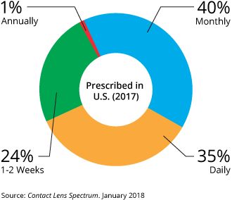
The most frequently prescribed contact lens replacement schedule in the U.S. in 2017 was monthly (40 percent), followed by daily (35 percent), every one to two weeks (24 percent) and annually (1 percent).

LEARN MORE about the proper care of your contact lenses.

Contact Lens Designs

Soft contact lenses (both standard hydrogel and silicone hydrogel lenses) are available in a variety of designs, depending on their intended purpose:

ExpandableSoft contact lens designs

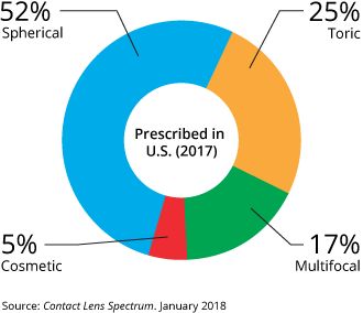
  • Spherical contact lenses have the same lens power throughout the entire optical part of the lens to correct myopia (nearsightedness) or hyperopia (farsightedness).
  • Toric soft contact lenses have different powers in different meridians of the lens to correct astigmatism as well as nearsightedness or farsightedness. [Read more about toric contact lenses.]
  • Multifocal contact lenses (including bifocal contacts) contain different power zones for near and far vision to correct presbyopia as well as nearsightedness or farsightedness. Some multifocal lenses also can correct astigmatism. [Read more about bifocal contacts.]
  • Cosmetic contact lenses include color contacts designed to change or intensify your eye color. Halloween, theatrical and other special-effect contacts also are considered cosmetic lenses. A contact lens prescription is required for cosmetic contacts even if you have no refractive errors that need correction.

All of these lenses can be custom made for hard-to-fit eyes. Other lens designs also are available — including lenses fabricated for use in special situations, such as correcting for keratoconus.

More Contact Lens Features

Bifocal contacts for astigmatism. These are advanced soft contacts that correct both presbyopia and astigmatism, so you can remain glasses-free after age 40 even if you have astigmatism. [More about bifocal contact lenses for astigmatism.]

Contacts for dry eyes. Are your contacts uncomfortably dry? Certain soft contact lenses are specially made to reduce the risk of contact lens-related dry eye symptoms. [More about contact lenses for dry eyes.]

Colored lenses. Many of the types of lenses described above also come in colors that can enhance the natural color of your eyes — that is, make your green eyes even greener, for example. Other colored lenses can totally change the color of your eyes, as in from brown to blue.

Special-effect lenses. Also called theatrical, novelty, or costume lenses, special-effect contacts take coloration one step further to make you look like a cat, a vampire, or another alter-ego of your choice.

Prosthetic lenses. Colored contact lenses also can be used for more medically oriented purposes. Opaque soft lenses called prosthetic contacts can be custom-designed for an eye that has been disfigured by injury or disease to mask the disfigurement and match the appearance of the other, unaffected eye.

Custom lenses. If conventional contact lenses don’t seem to work for you, you might be a candidate for custom contact lenses that are made-to-order for your individual eye shape and visual needs.

UV-inhibiting lenses. Some soft contact lenses help protect your eyes from the sun’s ultraviolet rays that can cause cataracts and other eye problems. But because contacts don’t cover your entire eye, you still should wear UV-blocking sunglasses outdoors for the best protection from the sun.

Scleral lenses. Large-diameter gas permeable lenses called scleral contacts are specially designed to treat keratoconus and other corneal irregularities, as well as presbyopia.

Myopia control contacts. Special contact lenses are being developed to slow or stop the progression of nearsightedness in children. [More about myopia control.]

Which Contact Lens Is Right for You?

First, your contacts must address the problem that is prompting you to wear lenses in the first place. Your contact lenses must provide good vision by correcting your myopiahyperopiaastigmatism, or some combination of those vision problems.

Second, the lens must fit your eye. To do that, lenses come in tens of thousands of combinations of diameter and curvature. Of course, not every lens brand comes in every “size.”

Your ECP is skilled in evaluating your eye’s physiology, and your eyesight, to determine which lens best satisfies the two criteria above.

Third, you may have another medical need that drives the choice of lens. For example, your ECP might pick a particular lens if your eyes tend to be dry.

Finally, consider your “wish list” of contact lens features — colors, for example, or overnight wear.

When you and your ECP decide on the right lens for you, you’ll be given a contact lens prescription. You’ll be able to buy a supply of lenses from your ECP or from the many other outlets that sell contact lenses. [For more on this, please read “Where’s the Best Place to Buy Contact Lenses?”]

Contact Lens Wear and Care

Caring for your contact lenses — cleaning, disinfecting and storing them — is much easier than it used to be.

A few years ago, you would have needed several bottles of cleaning products, and perhaps enzyme tablets, for proper care. Today, most people can use “multipurpose” solutions — meaning that one product both cleans and disinfects, and is used for storage.

People who are sensitive to the preservatives in multipurpose solutions might need preservative-free systems, such as those containing hydrogen peroxide. These do an excellent job of cleaning contacts, but it’s very important to follow the directions for using them. The solution should not come into contact with your eyes until soaking is complete and the solution is neutralized.

Of course, you can avoid lens care altogether by wearing daily disposable contact lenses.

Contact Lens Problems

Trial and error often is involved in finding the perfect lens for you. People react differently to various lens materials and cleaning solutions.

Also, the correct “parameters” of your lens — that is, power, diameter, and curvature — can be finalized only after you’ve successfully worn the lens. This is especially true for more complex fits involving extra parameters, such as with bifocals or toric contact lenses for astigmatism.

If you experience discomfort or poor vision when wearing contact lenses, chances are that an adjustment or change of lens can help.

Today, more contact lens choices than ever are available to provide comfort, good vision, and healthy eyes. If your eyes or lenses are uncomfortable or you are not seeing well, remove your lenses and visit your eye care professional to explore available remedies for contact lens discomfort.

Buying Contact Lenses

You can buy replacement contact lenses at many places, and some offer a better value than others. Find out more about where to buy contacts and buying contact lenses online.

More Information About Contacts

For more information about contacts, visit our contact lens expert at Eyeupdate Eye clinic, 01 Ajuwon junction beside BPNL Filling station, Akute/Ajuwon road, Off Elliot bus stop, Iju-Ishaga, Lagos/Ogun


Contact lenses & solution

Buy prescriptive contact lenses @ N5000 per pair with solution and storage case. Call 08034971582 to place your order

Eye care tips – Eye/Optical clinic near Obawole Fagba Iju Ishaga

Your eyes are an important part of your health. Most people rely on their eyes to see and make sense of the world around them. But some eye diseases can lead to vision loss, so it is important to identify and treat eye diseases as early as possible. You should get your eyes checked as often as your health care provider recommends it, or if you have any new vision problems. And just as it is important to keep your body healthy, you also need to keep your eyes healthy.

Eye Care Tips

There are things you can do to help keep your eyes healthy and make sure you are seeing your best:

  • Eat a healthy, balanced diet. Your diet should include plenty or fruits and vegetables, especially deep yellow and green leafy vegetables. Eating fish high in omega-3 fatty acids, such as salmon, tuna, and halibut can also help your eyes.
  • Maintain a healthy weight. Being overweight or having obesity increases your risk of developing diabetes. Having diabetes puts you at higher risk of getting diabetic retinopathy or glaucoma.
  • Get regular exercise. Exercise may help to prevent or control diabetes, high blood pressure, and high cholesterol. These diseases can lead to some eye or vision problems. So if you exercise regularly, you can lower your risk of getting these eye and vision problems.
  • Wear Transition lenses. Sun exposure can damage your eyes and raise your risk of cataracts and age-related macular degeneration. Protect your eyes by using sunglasses that block out 99 to 100 percent of both UV-A and UV-B radiation.
  • Wear protective eye wear. To prevent eye injuries, you need eye protection when playing certain sports, working in jobs such as factory work and construction, and doing repairs or projects in your home.
  • Avoid smoking. Smoking increases the risk of developing age-related eye diseases such as macular degeneration and cataracts and can damage the optic nerve.
  • Know your family medical history. Some eye diseases are inherited, so it is important to find out whether anyone in your family has had them. This can help you determine if you are at higher risk of developing an eye disease.
  • Know your other risk factors. As you get older, you are at higher risk of developing age-related eye diseases and conditions. It is important to know you risk factors because you may be able to lower your risk by changing some behaviors.
  • If you wear contacts, take steps to prevent eye infections. Wash your hands well before you put in or take out your contact lenses. Also follow the instructions on how to properly clean them, and replace them when needed.
  • Give your eyes a rest. If you spend a lot of time using a computer, you can forget to blink your eyes and your eyes can get tired. To reduce eyestrain, try the 20-20-20 rule: Every 20 minutes, look away about 20 feet in front of you for 20 seconds.

Eye Tests and Exams

Everyone needs to have their eyesight tested to check for vision and eye problems. Children usually have vision screening in school or at their health care provider’s office during a checkup. Adults may also get vision screenings during their checkups. But many adults need more than a vision screening. They need a comprehensive dilated eye exam.

Getting comprehensive dilated eye exams is especially important because some eye diseases may not have warning signs. The exams are the only way to detect these diseases in their early stages, when they are easier to treat.

The exam includes several tests:

  • A visual field test to measure your side (peripheral) vision. A loss of peripheral vision may be a sign of glaucoma.
  • A visual acuity test, where you read an eye chart about 20 feet away, to check on how well you see at various distances
  • Tonometry, which measures your eye’s interior pressure. It helps to detect glaucoma.
  • Dilation, which involves getting eye drops that dilate (widen) your pupils. This allows more light to enter the eye. Your eye care provider examines your eyes using a special magnifying lens. This provides a clear view of important tissues at the back of your eye, including the retina, macula, and optic nerve.

If you have a refractive error and are going to need glasses or contacts, then you will also have a refraction test. When you have this test, you look through a device that has lenses of different strengths to help your eye care professional figure out which lenses will give you the clearest vision.

At what age you should start getting these exams and how often you need them depends on many factors. They include your age, race, and overall health. For example, if you are African American, you are at higher risk of glaucoma and you need to start getting the exams earlier. If you have diabetes, you should get an exam every year. Check with your health care provider about if and when you need these exams

Eye test – Eye clinic within Obawole Iju Ishagah Fagba

Dr. Dr. Steven E.N., Eyeupdate clinic & optical supplies, 01 Ajuwon junction, beside BPNL filling station, Ajuwon, off Elliot bus stop, Iju-Ishagah, Lagos. Tel: 08107531046, 08034971582.


Eye exam/test consists of series of tests that your eye doctor will perform in order to determine your ocular status.

An eye exam involves a series of tests to evaluate your vision and check for eye diseases/vision defects. Your eye doctor may use a variety of instruments, shine bright lights directly at your eyes and request that you look through an array of lenses. Each test during an eye exam evaluates a different aspect of your vision or eye health.

Why it’s done

An eye exam helps detect eye problems at their earliest stage — when they’re most treatable. Regular eye exams give your eye care professional a chance to help you correct or adapt to vision changes and provide you with tips on caring for your eyes.

When to have an eye exam

Several factors may determine how frequently you need an eye exam, including your age, health and risk of developing eye problems. General guidelines are as follows:

Children 3 years and younger

For children under 3, your pediatrician will likely look for the most common eye problems — lazy eye, crossed eyes or misaligned eyes. If there are eye concerns or symptoms, an examination is appropriate at that time regardless of age. Your child could undergo a more comprehensive eye exam between the ages of 3 and 5.

School-age children and adolescents

Have your child’s vision checked before he or she enters first grade. If your child has no symptoms of vision problems and no family history of vision problems, have his or her vision rechecked every one to two years. Otherwise, schedule eye exams based on the advice of your eye doctor.

Adults

In general, if you are healthy and you have no symptoms of vision problems, have your eyes checked on this schedule:

  • Every five to 10 years in your 20s and 30s
  • Every two to four years from 40 to 54
  • Every one to three years from 55 to 64
  • Every one to two years after age 65

Have your eyes checked more often if you:

  • Wear glasses or contact lenses
  • Have a family history of eye disease or loss of vision
  • Have a chronic disease that puts you at greater risk of eye disease, such as diabetes
  • Take medications that have serious eye side effects
  • How you prepare

    There are three different types of eye specialists. Which specialist you choose may be a matter of personal preference or will depend on the nature of your eye problem.

    • Ophthalmologists. Ophthalmologists are medical doctors who provide full eye care, such as giving you a complete eye exam, prescribing corrective lenses, diagnosing and treating complex eye diseases, and performing eye surgery.
    • Optometrists. Optometrists provide many of the same services as ophthalmologists, such as evaluating your vision, prescribing corrective lenses, diagnosing common eye disorders and treating selected eye diseases with drugs. If you have a complex eye problem or need surgery, your doctor can refer you to an ophthalmologist.
    • Opticians. Opticians fill prescriptions for eyeglasses, including assembling, fitting and selling them. Some opticians also sell contact lenses. Opticians do not provide eye health evaluations.

    Bring your prescription eyewear

    If you wear contact lenses or glasses, bring them to your appointment. Your eye doctor will want to make sure your prescription is the best one for you.

    Other precautions

    If your eyes are dilated as a part of your eye exam, you may want to bring sunglasses to wear after your eye exam is complete, as daylight or other bright lights may be uncomfortable or cause blurred vision. Also, consider having someone else drive you home.

    What you can expect

    Before the exam

    If you’re seeing a new eye doctor or if you’re having your first eye exam, expect questions about your vision history. Part of the examination, such as taking your medical history and the initial eye test, may be performed by a clinical assistant or technician.

    Your answers help your eye doctor understand your risk of eye disease and vision problems. Be prepared to give specific information, including:

    • Are you having any eye problems now?
    • Have you had any eye problems in the past?
    • Do you wear glasses or contacts now? If so, are you satisfied with them?
    • What health problems have you had in recent years?
    • Were you born prematurely?
    • Are you taking any medications?
    • Do you have any allergies to medications, food or other substances?
    • Have you ever had eye surgery?
    • Does anyone in your family have eye problems, such as macular degeneration, glaucoma or retinal detachments?
    • Do you or does anyone in your family have diabetes, high blood pressure, heart disease or any other health problems that can affect the whole body?

    During the exam

    An eye exam usually involves these steps:

    • You’ll be asked about your medical history and any vision problems you might be experiencing.
    • Your eye doctor measures your visual acuity to see if you need glasses or contact lenses to improve your vision.
    • You’ll be given a numbing drop in your eyes. Then your eye pressure is measured. To make it easier for your doctor to examine the inside of your eye, he or she will likely dilate your eyes with eyedrops.
    • After waiting for the dilating drops to take effect, your eye doctor checks the health of your eyes, possibly using several lights to evaluate the front of the eye and the inside of each eye.

    Several different tests may be performed during the eye exam. The tests are designed to check your vision and to examine the appearance and function of all parts of your eyes.

    After the exam

    At the end of your eye exam, you and your doctor will discuss the results of all testing, including an assessment of your vision, your risk of eye disease and preventive measures you can take to protect your eyesight.

    Different types of eye exams

    Eye muscle test

    This test evaluates the muscles that control eye movement. Your eye doctor watches your eye movements as you follow a moving object, such as a pen or small light, with your eyes. He or she looks for muscle weakness, poor control or poor coordination.

    Visual acuity test

    This test measures how clearly you see. Your doctor asks you to identify different letters of the alphabet printed on a chart (Snellen chart) or a screen positioned some distance away. The lines of type get smaller as you move down the chart. Each eye is tested separately. Your near vision also may be tested, using a card with letters similar to the distant eye chart. The card is held at reading distance.

    Refraction assessment

    Light waves are bent as they pass through your cornea and lens. If light rays don’t focus perfectly on the back of your eye, you have a refractive error. Having a refractive error may mean you need some form of correction, such as glasses, contact lenses or refractive surgery, to see as clearly as possible.

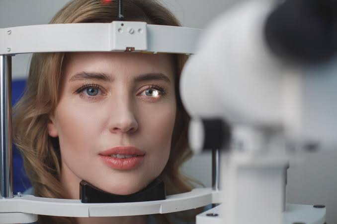
    Assessment of your refractive error helps your doctor determine a lens prescription that will give you the sharpest, most comfortable vision. The assessment may also determine that you don’t need corrective lenses.

    Your doctor may use a computerized refractor to estimate your prescription for glasses or contact lenses. Or he or she may use a technique called retinoscopy. In this procedure, the doctor shines a light into your eye and measures the refractive error by evaluating the movement of the light reflected by your retina back through your pupil.

    Your eye doctor usually fine-tunes this refraction assessment by having you look through a masklike device that contains wheels of different lenses (phoropter). He or she asks you to judge which combination of lenses gives you the sharpest vision.

    Visual field test (perimetry)

    Your visual field is the full extent of what you can see to the sides without moving your eyes. The visual field test determines whether you have difficulty seeing in any areas of your overall field of vision. The different types of visual field tests include:

    • Confrontation exam. Your eye doctor sits directly in front of you and asks you to cover one eye. You look straight ahead and tell the doctor each time you see his or her hand move into view.
    • Manual testing, including tangent screen and Goldmann exams. You sit a short distance from a screen and focus on a target at its center. You tell the doctor when you can see an object move into your peripheral vision and when it disappears.
    • Automated perimetry. As you look at a screen with blinking lights on it, you press a button each time you see a blink.

    Using your responses to one or more of these tests, your eye doctor determines the fullness of your field of vision. If you aren’t able to see in certain areas, noting the pattern of your visual field loss may help your eye doctor diagnose your eye condition.

    Color vision testing

    You could have poor color vision and not even realize it. If you have difficulty distinguishing certain colors, your eye doctor may screen your vision for a color deficiency. To do this, your doctor shows you several multicolored dot-pattern tests.

    If you have no color deficiency, you’ll be able to pick out numbers and shapes from within the dot patterns. If you do have a color deficiency, you’ll find it difficult to see certain patterns within the dots. Your doctor may use other tests, as well.

    Slit-lamp examination

    A slit lamp is a microscope that magnifies and illuminates the front of your eye with an intense line of light. Your doctor uses this device to examine the eyelids, lashes, cornea, iris, lens and fluid chamber between your cornea and iris.

    Your doctor may use a dye, most commonly fluorescein (flooh-RES-een), to color the film of tears over your eye. This helps reveal any damaged cells on the front of your eye. Your tears wash the dye from the surface of your eye fairly quickly.

    Retinal examination

    A retinal examination — sometimes called ophthalmoscopy or funduscopy — allows your doctor to evaluate the back of your eye, including the retina, the optic disk and the underlying layer of blood vessels that nourish the retina (choroid). Usually before your doctor can see these structures, your pupils must be dilated with eyedrops that keep the pupil from getting smaller when your doctor shines light into the eye.

    After administering eyedrops and giving them time to work, your eye doctor may use one or more of these techniques to view the back of your eye:

    • Direct exam. Your eye doctor uses an ophthalmoscope to shine a beam of light through your pupil to see the back of the eye. Sometimes eyedrops aren’t necessary to dilate your eyes before this exam.
    • Indirect exam. During this exam, you might lie down, recline in a chair or sit up. Your eye doctor examines the inside of the eye with the aid of a condensing lens and a bright light mounted on his or her forehead. This exam lets your doctor see the retina and other structures inside your eye in great detail and in three dimensions.

    Screening for glaucoma

    Tonometry measures the fluid pressure inside your eye (intraocular pressure). This is one test that helps your eye doctor detect glaucoma, a disease that damages the optic nerve.

    Several methods to measure intraocular pressure are available, including:

    • Applanation tonometry. This test measures the amount of force needed to temporarily flatten a part of your cornea. You’ll be given eyedrops with fluorescein, the same dye used in a regular slit-lamp examination. You’ll also receive eyedrops containing an anesthetic. Using the slit lamp, your doctor moves the tonometer to touch your cornea and determine the eye pressure. Because your eye is numbed, the test doesn’t hurt.
    • Noncontact tonometry. This method uses a puff of air to estimate the pressure in your eye. No instruments touch your eye, so you won’t need an anesthetic. You’ll feel a momentary pulse of air on your eye, which can be startling.

    If your eye pressure is higher than average or your optic nerve looks unusual, your doctor may use a pachometer. This instrument uses sound waves to measure the thickness of your cornea. The most common way of measuring corneal thickness is to put an anesthetic drop in your eye, then place a small probe in contact with the front surface of the eye. The measurement takes seconds.

    You may need more-specialized tests, depending on your age, medical history and risk of developing eye disease.

    Results

    Normal results from an eye exam include:

    • 20/20 vision
    • Good peripheral vision
    • Ability to distinguish various colors
    • Normal-appearing structures of the external eye
    • Absence of cataract, glaucoma or retinal disorders, such as macular degeneration

    Your doctor may give you a prescription for corrective lenses. If your eye exam yields other abnormal results, your doctor will discuss with you next steps for further testing or for treating an underlying condition.

High blood pressure & your eyes – Eye clinic near Sango Ota Abule Egba and Fagba

Smoking and your eyes – Eye clinic near Mowe Ojodu Berger

Eye Disease and Smoking:

Smoking has long been known to cause heart disease and lung cancer; however many people don’t realize that smoking can lead to vision loss. Studies show smoking increases the risk of age-related macular degeneration, cataracts, glaucoma and diabetic retinopathy and Dry Eye Syndrome.

  • Age Related Macular Degeneration (AMD)
    • One way to reduce the risk of developing AMD is by NOT smoking. Smokers are three to four times more likely to develop AMD than nonsmokers. Nonsmokers living with smokers almost double their risk of developing AMD.
  • Cataract
    • Heavy smokers (15 cigarettes/day or more) have up to three times the risk of cataract as nonsmokers.
  • Glaucoma
    • There is a strong link between smoking and high blood pressure, cataracts and diabetes all of which are risk factors for glaucoma.
  • Diabetic Retinopathy
    • Smoking can increase your chances of getting diabetes. It can also make managing diabetes more difficult for those who already have it. Complications of diabetes made worse by smoking include retinopathy, heart disease, stroke, vascular disease, kidney disease, nerve damage, foot problems and many others.
  • Dry Eye Syndrome
    • Dry Eye Syndrome is more than twice as likely to impact smokers as non-smokers.

What You Can Do to Prevent Vision Loss:

Healthy habits can lead to healthy eyes. The risk of eye disease and vision loss can be lowered if you::

  • Quit smoking!
  • Eat healthy foods (including green leafy vegetables, fruits and foods high in vitamins C, E, and beta carotene).
  • Control blood pressure and cholesterol.
  • Stay active.
  • Visit your eye care professional regularly.

Are You Ready to Quit?

  • To get started, visit the How to Quit page on the NYS Smokers’ Quitline website ( www.nysmokefree.com ) or call the NYS Smokers’ Quitline for help <( 1-866-NY-QUITS or 1-866-697-8487).
  • See your doctor. He or she may prescribe a nicotine replacement therapy or other medication.

Description of Eye Diseases Associated with Smoking:

1. Age Related Macular Degeneration (AMD):

AMD begins as a loss of central vision which makes it difficult to read and see fine details. Over time, vision loss increases significantly. Of the two types of AMD, “dry” and “wet,” dry AMD is the most common. In dry AMD, fatty deposits form under the light-sensing cells in the back of the eye (retina). Vision loss in dry AMD usually gets worse slowly. In wet AMD, tiny blood vessels under the retina leak or break open. This changes vision and causes scar tissue to form. Wet AMD is less common, but more quickly harmful to vision.

2. Glaucoma

Glaucoma causes a gradual break down of the cells that make up the nerve in your eye that sends visual information to your brain (optic nerve). As the nerve cells die, vision is slowly lost, usually beginning with side vision. Often the loss of vision is not noticeable until a large amount of nerve damage has occurred. This is the reason why as many as half of all people with glaucoma may be unaware that they have it.

3. Cataract

Cataract is a clouding of the eye’s naturally clear lens. It usually gets worse as we get older. Most cataracts are related to aging. Cataracts are very common in older people. By age 80, more than half of all Americans either have a cataract or have had cataract surgery.

4. Diabetic retinopathy

Diabetic retinopathy is a common complication of diabetes. It affects the tiny blood vessels of the retina in the eye. Retinal blood vessels can break down, leak or become blocked and this can affect vision over time. In some people with diabetic retinopathy, serious damage to the eye can occur when new blood vessels grow on the surface of the retina.

5. Dry Eye Syndrome

Dry Eye Syndrome is an eye disease that appears as damaged blood vessels in the eye. This can lead to eye irritation, itchy and scratchy eyes, and burning sensation of the eyes.